


Topic
- Acromial end of clavicle
- Acromial part of deltoid muscle
- Acromioclavicular joint
- Acromioclavicular ligament
- Acromion process of scapula
- Adipose tissue (Shoulder)
- Anatomical neck of humerus
- Anterior glenoid labrum
- Anterior rim of glenoid
- Articular cartilage of glenoid fossa
- Axillary artery
- Axillary lymph nodes
- Axillary nerve
- Biceps brachii muscle
- Biceps pulley
- Bicipital groove
- Body of humerus
- Body of scapula
- Brachial artery
- Brachial plexus
- Cephalic vein
- Circumflex scapular artery
- Circumflex subscapular artery
- Clavicle
- Clavicular part of deltoid muscle
- Conoid ligament
- Conoid tubercle
- Coracoacromial ligament
- Coracobrachialis muscle
- Coracoclavicular Ligaments
- Coracohumeral ligament
- Coracoid process of scapula
- Crest of greater tubercle
- Crest of lesser tubercle
- Deltoid Tendon (Proximal)
- Deltoid muscle
- Deltoid tendon (Distal)
- Dorsal scapular artery
- Dorsal scapular nerve
- Glenohumeral joint capsule
- Glenohumeral ligaments
- Glenoid fossa
- Glenoid labrum
- Glenoid process of scapula
- Greater tubercle of humerus
- Head of humerus
- Humerus
- Inferior acromioclavicular ligament
- Inferior angle of scapula
- Inferior belly of omohyoid muscle
- Inferior glenohumeral ligament
- Inferior glenohumeral ligament anterior band
- Inferior glenohumeral ligament posterior band
- Inferior glenohumeral ligament, axillary pouch
- Inferior transverse scapular ligament
- Infraglenoid tubercle
- Infraspinatus muscle
- Infraspinatus tendon
- Infraspinous fossa
- Lateral border of scapula
- Lateral head of triceps brachii muscle
- Latissimus dorsi tendon
- Left common carotid artery
- Left lung
- Lesser tubercle of humerus
- Levator scapulae muscle
- Long head of biceps brachii muscle
- Long head of biceps tendon
- Long head of triceps brachii muscle
- Long thoracic nerve
- Medial border of scapula
- Medial cutaneous nerve of forearm
- Medial head of triceps brachii muscle
- Median nerve
- Middle glenohumeral ligament
- Neck of scapula
- Pectoralis major muscle
- Pectoralis minor muscle
- Posterior circumflex humeral artery
- Posterior circumflex humeral vein
- Posterior cutaneous nerve of arm
- Posterior glenoid labrum
- Posterior rim of glenoid
- Radial nerve
- Ribs
- Scapula
- Scapular body
- Scapular spinal part of deltoid muscle
- Shaft (body) of clavicle
- Shaft of humerus
- Short head of biceps brachii muscle
- Short head of the biceps brachii tendon
- Shoulder joint (glenohumeral joint)
- Spine of scapula
- Spiral glenohumeral ligament
- Sternoclavicular joint
- Subacromial space
- Subclavius muscle
- Subscapular artery
- Subscapular fossa
- Subscapularis muscle
- Subscapularis tendon
- Superior acromioclavicular ligament
- Superior angle of scapula
- Superior border of scapula
- Superior glenohumeral ligament
- Superior transverse scapular ligament
- Supraclavicular fossa
- Supraglenoid tubercle
- Suprascapular artery
- Supraspinatus muscle
- Supraspinatus tendon
- Surgical neck of humerus
- Teres major muscle
- Teres major tendon (Distal)
- Teres minor muscle
- Teres minor tendon (Distal)
- Thoracodorsal artery
- Transverse cervical artery
- Transverse humeral ligament
- Trapezoid Line
- Trapezoid ligament
- Triceps brachii muscle
- Tuberosity of ulna
- Ulnar nerve
- supraspinous fossa of scapula
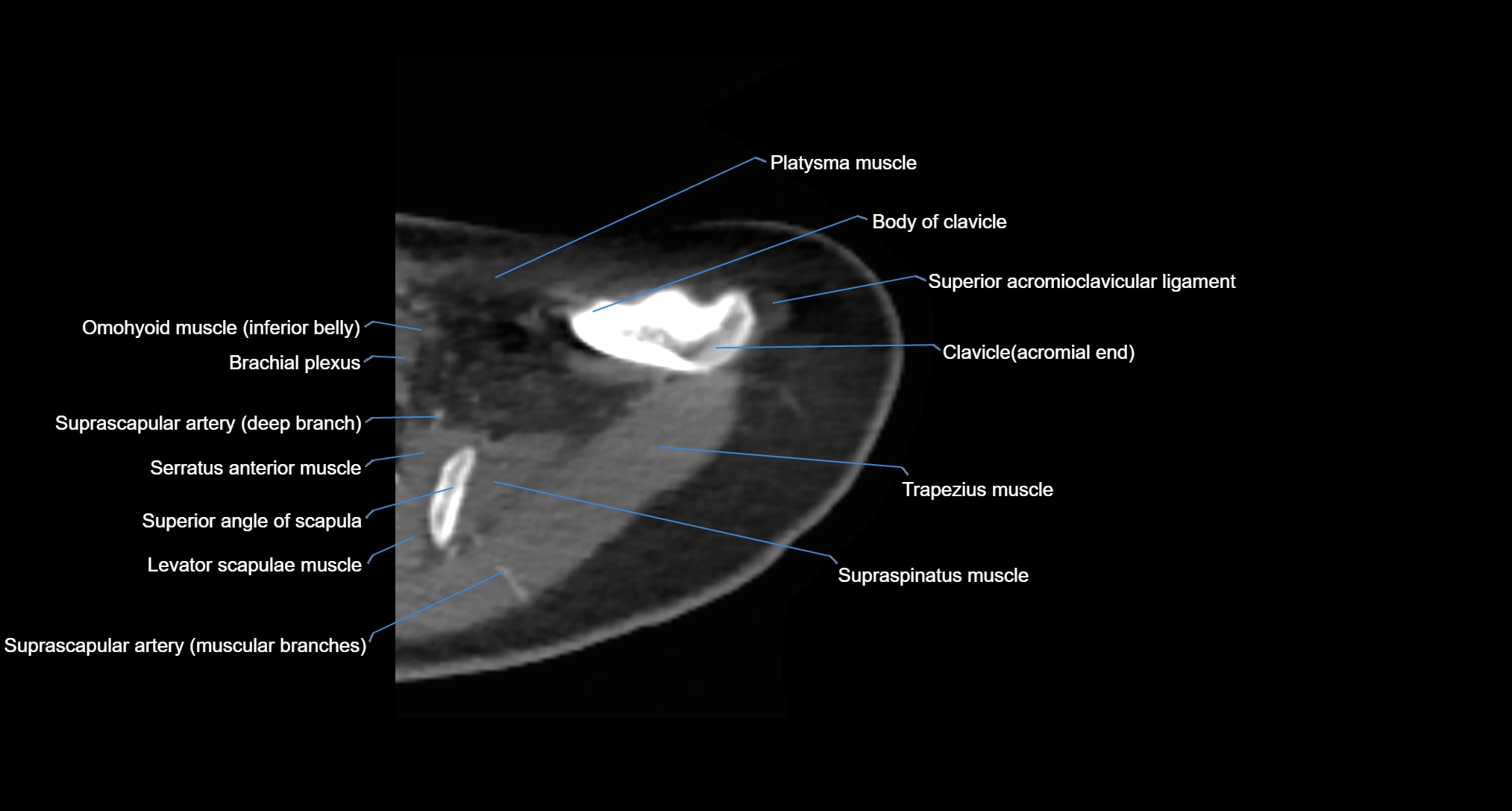
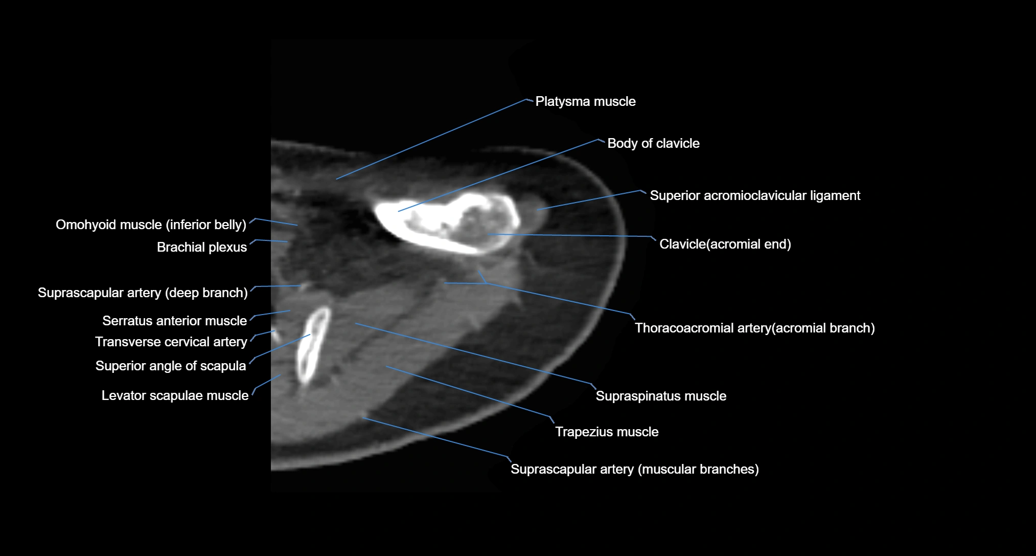
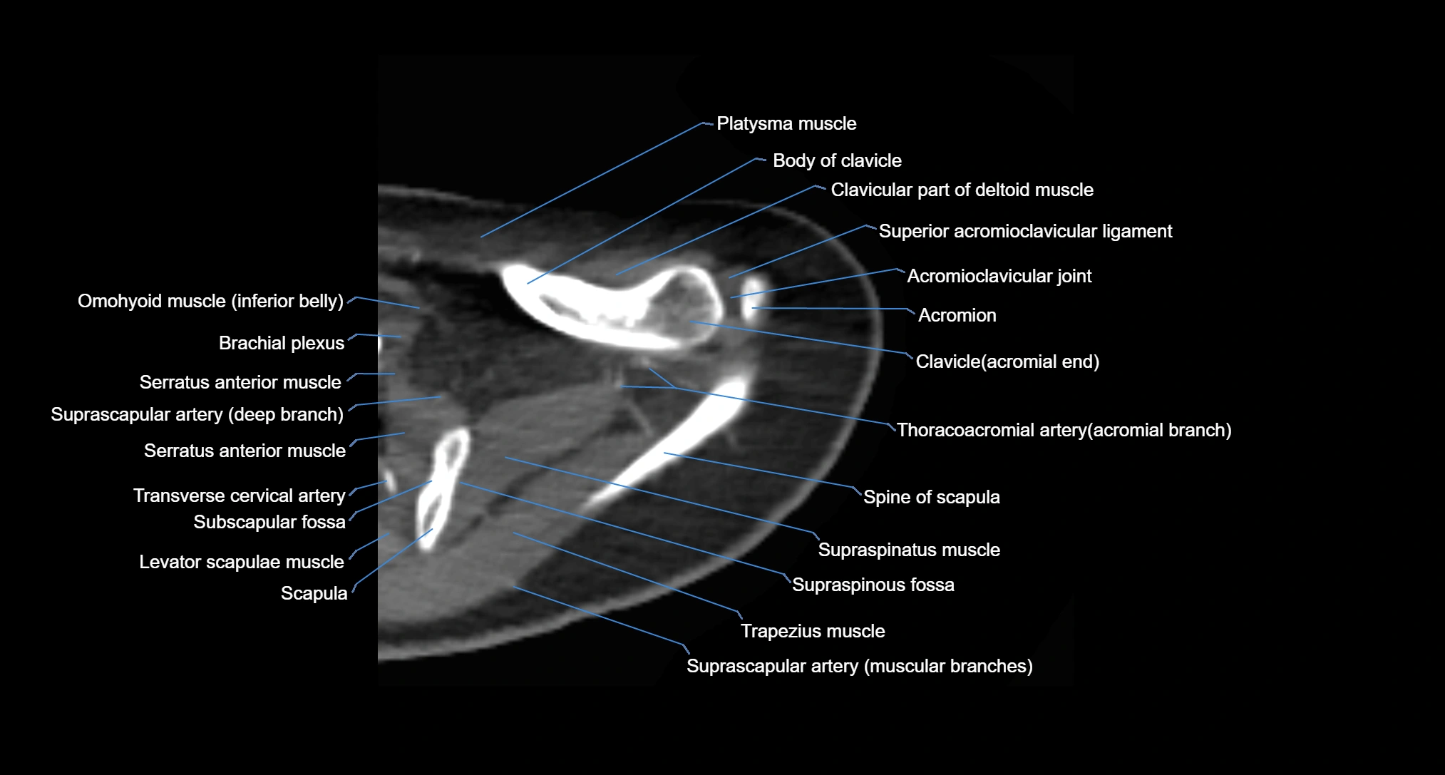
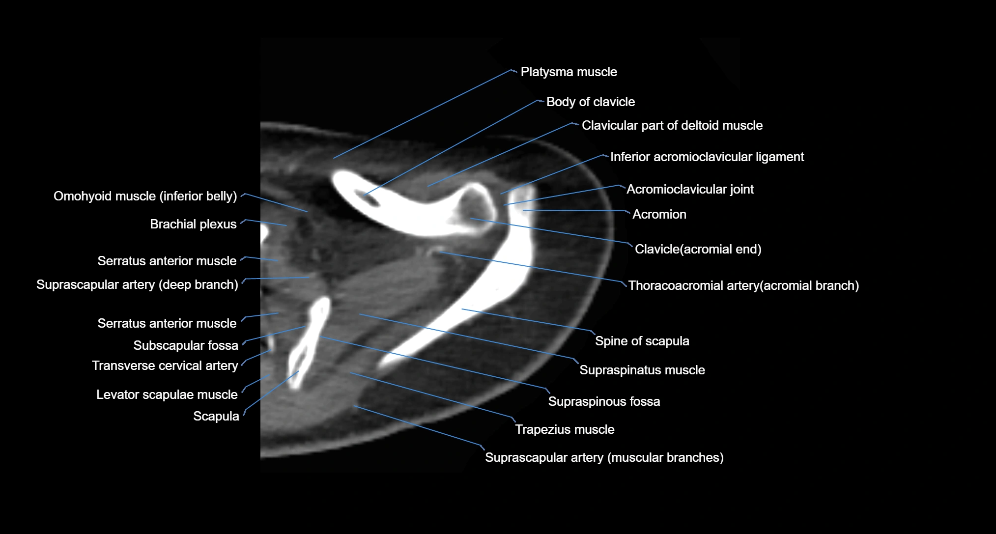
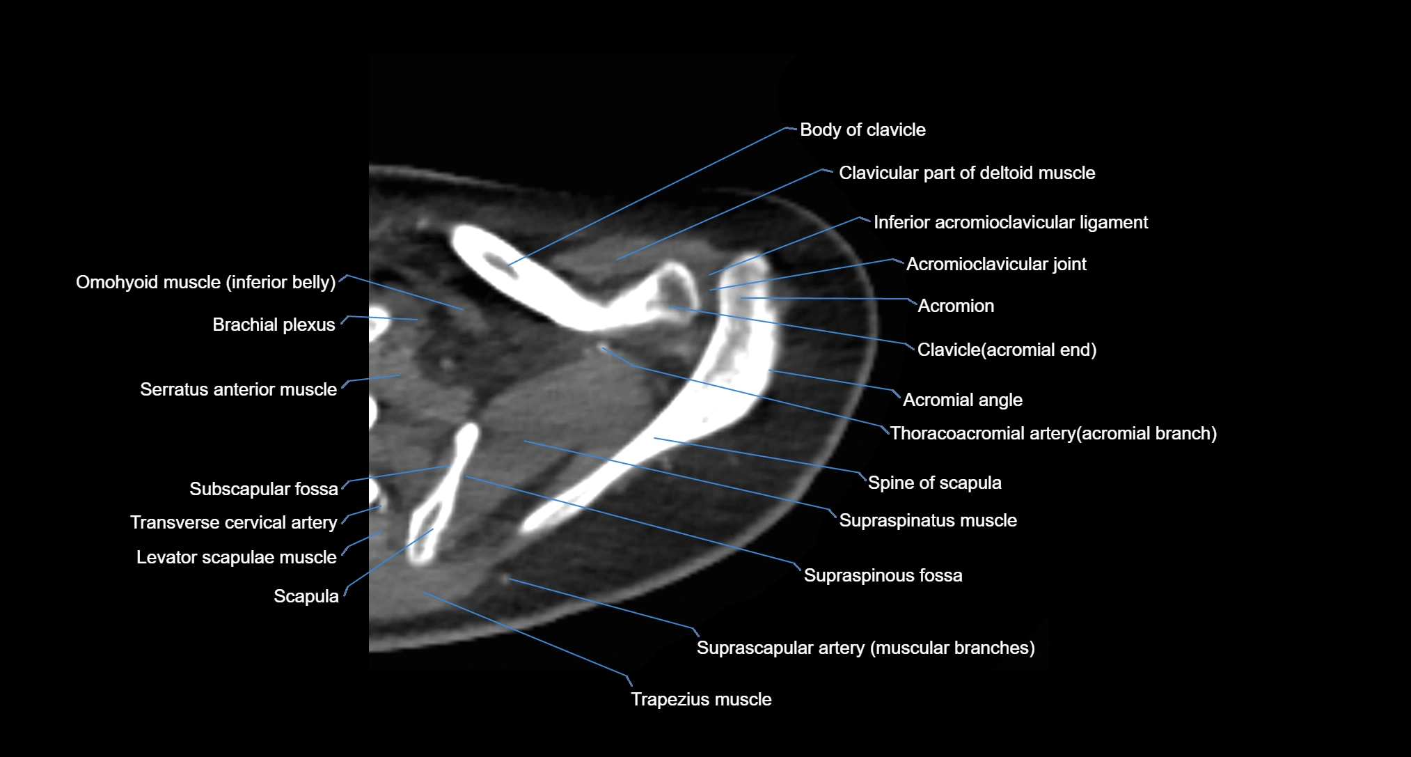
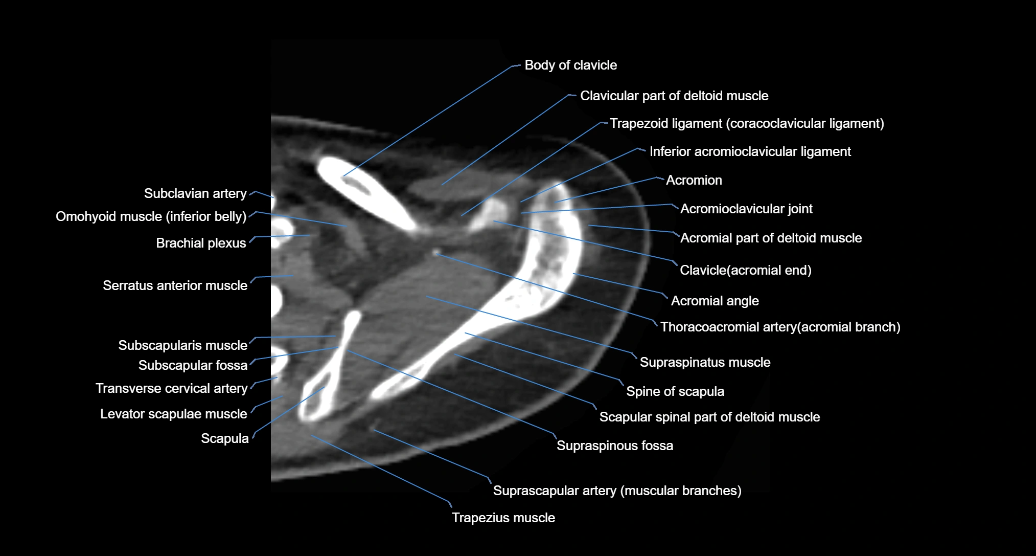
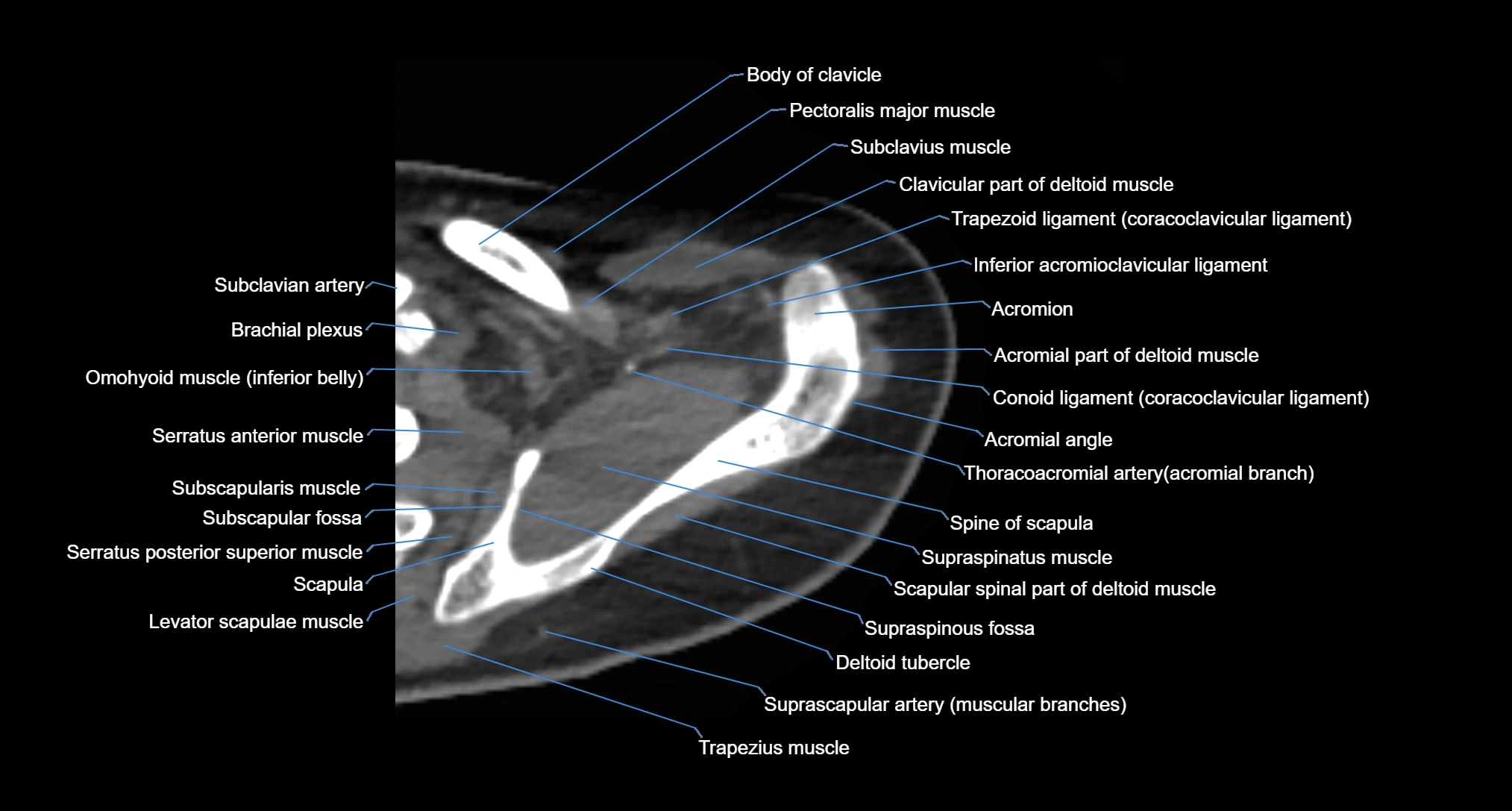
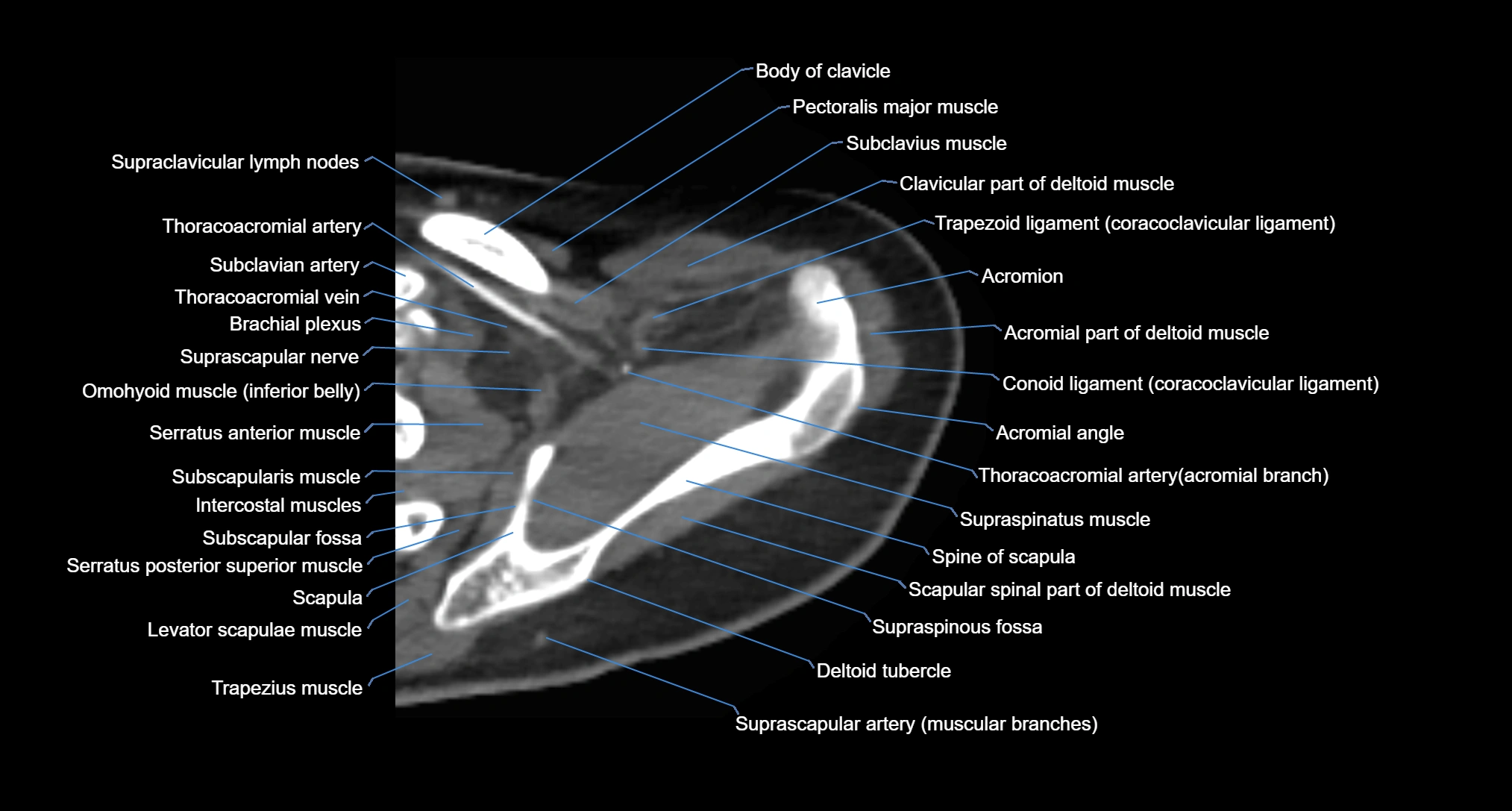
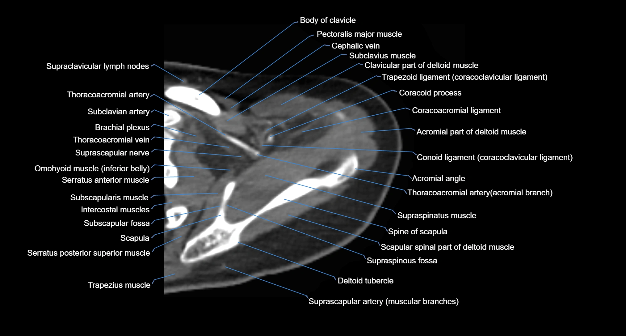
The acromial end of the clavicle is the flattened lateral extremity of the clavicle that articulates with the acromion of the scapula to form the acromioclavicular (AC) joint. Unlike the sternal end, the acromial end is broad and compressed. Its articular surface is oval, directed downward and medially, and covered with fibrocartilage.
The AC joint is stabilized by the acromioclavicular ligaments (superior and inferior) and reinforced by the coracoclavicular ligaments (conoid and trapezoid), which prevent vertical displacement. Small intra-articular fibrocartilaginous discs may be present.
This region is highly mobile, allowing scapular rotation, gliding, and elevation, which are essential for full shoulder motion. It is clinically significant as a frequent site of degeneration, separation injuries, fractures, and osteoarthritis.
Synonyms
-
Lateral end of clavicle
-
Acromioclavicular end
-
Acromial extremity of clavicle
Function
-
Forms the AC joint, connecting the clavicle and scapula
-
Transmits forces from the upper limb to the axial skeleton
-
Provides attachment for the acromioclavicular and coracoclavicular ligaments
-
Enables scapular rotation and stability of the shoulder girdle
MRI Appearance
T1-weighted images:
-
Bone marrow: intermediate signal
-
Cortical bone: hypointense rim
-
AC joint space: visible as a thin hypointense line
T2-weighted images:
-
Cartilage: hyperintense
-
Joint fluid or effusion: bright signal
-
Detects degenerative changes and joint inflammation
PD-FS (Proton Density Fat-Suppressed):
-
Enhances visualization of capsule, ligaments, and marrow edema
-
AC joint pathology (arthritis, capsular injury, synovitis) appears hyperintense
-
Excellent for trauma and subtle instability assessment
STIR:
-
Suppresses fat, highlighting marrow edema and soft tissue inflammation
-
Useful in acute fractures and ligamentous injuries
T1 Post-Gadolinium (MR Arthrography or contrast-enhanced MRI):
-
Highlights capsule, synovium, and ligamentous insertions
-
Detects synovitis, capsular tears, and enhancing arthropathy
MRI Non-Contrast 3D Imaging:
-
Provides 3D reconstructions of joint morphology, acromial end alignment, and cartilage surface
-
Useful in pre-surgical planning for distal clavicle excision or AC reconstruction
CT Appearance
Non-contrast CT:
-
Best for cortical detail: fractures, erosions, bone spurs, and joint alignment
-
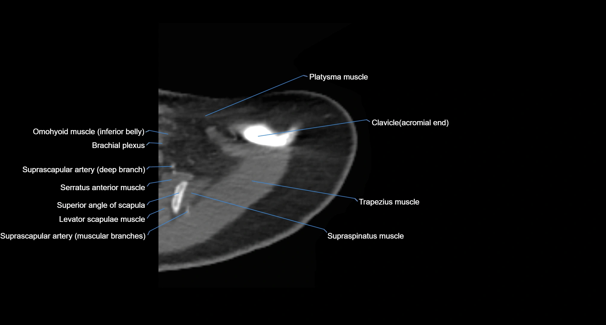
Defines the relationship of the acromial end with the acromion
CT Post-Contrast (CT Arthrography):
-
Contrast highlights the AC joint capsule and articular cartilage
-
Detects capsular tears, cartilage loss, osteoarthritis, and subtle osseous injury
-
3D reconstructions help in planning reconstructive surgery or distal clavicle excision
MRI images

MRI images

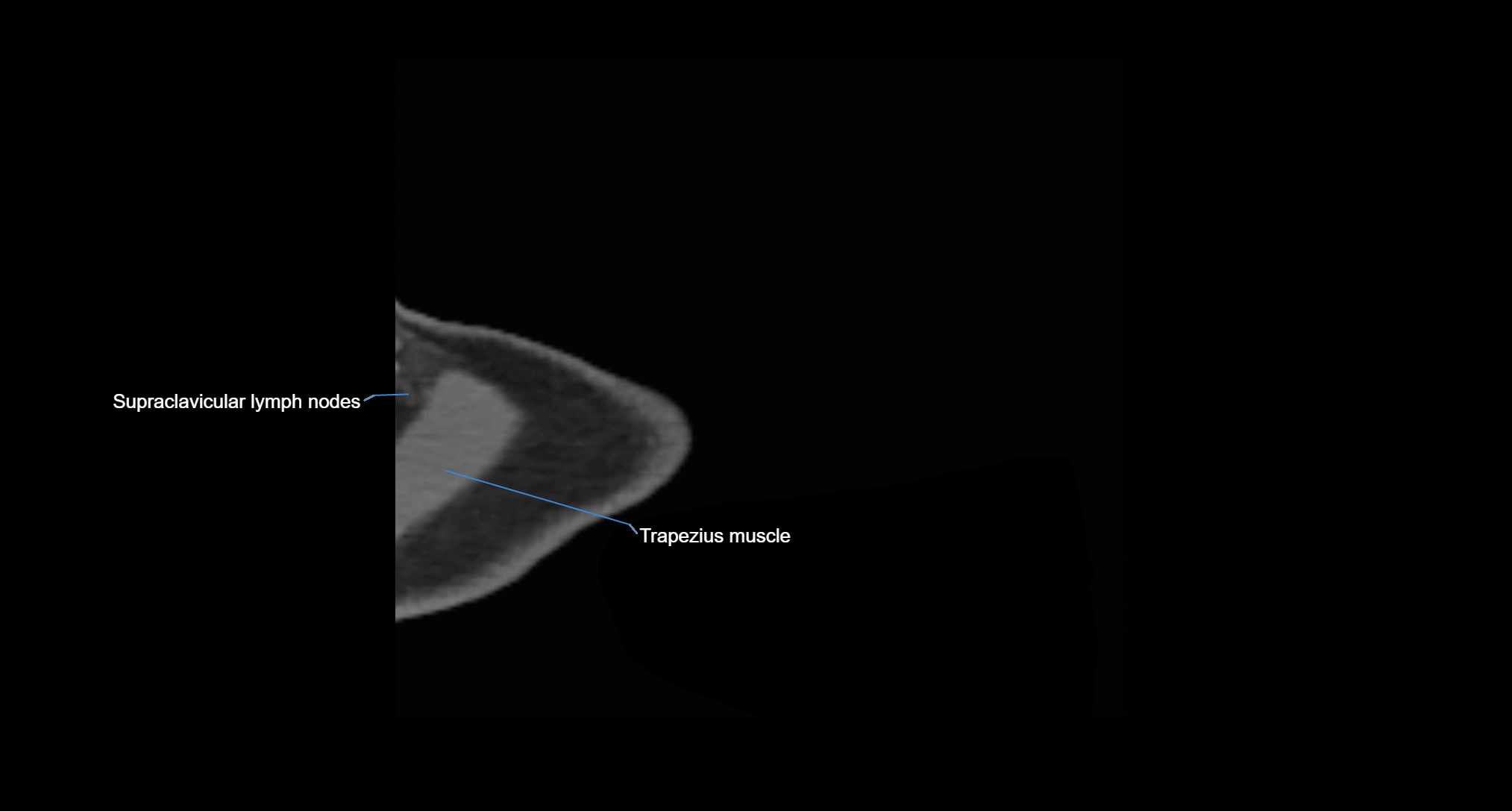
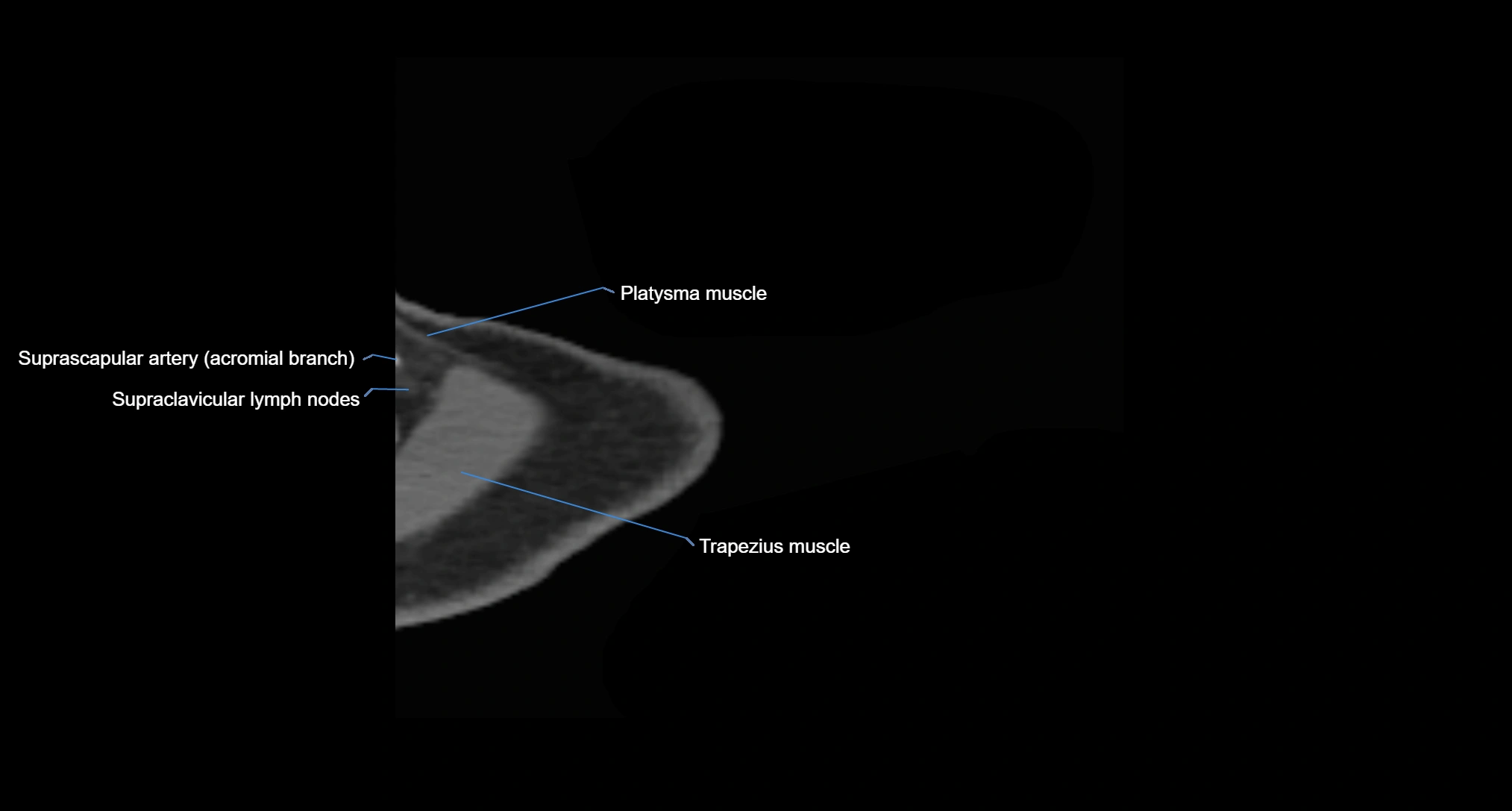
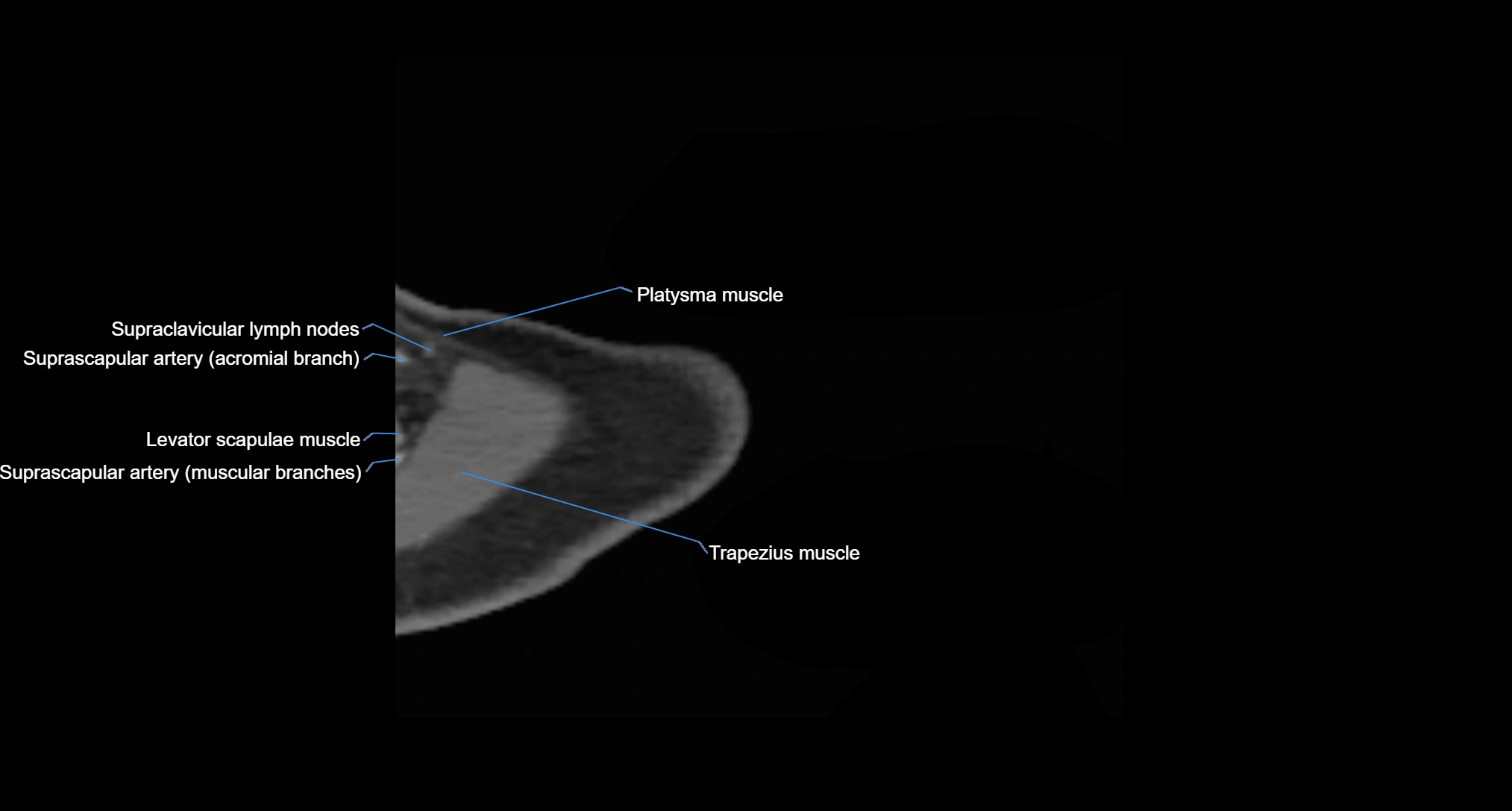
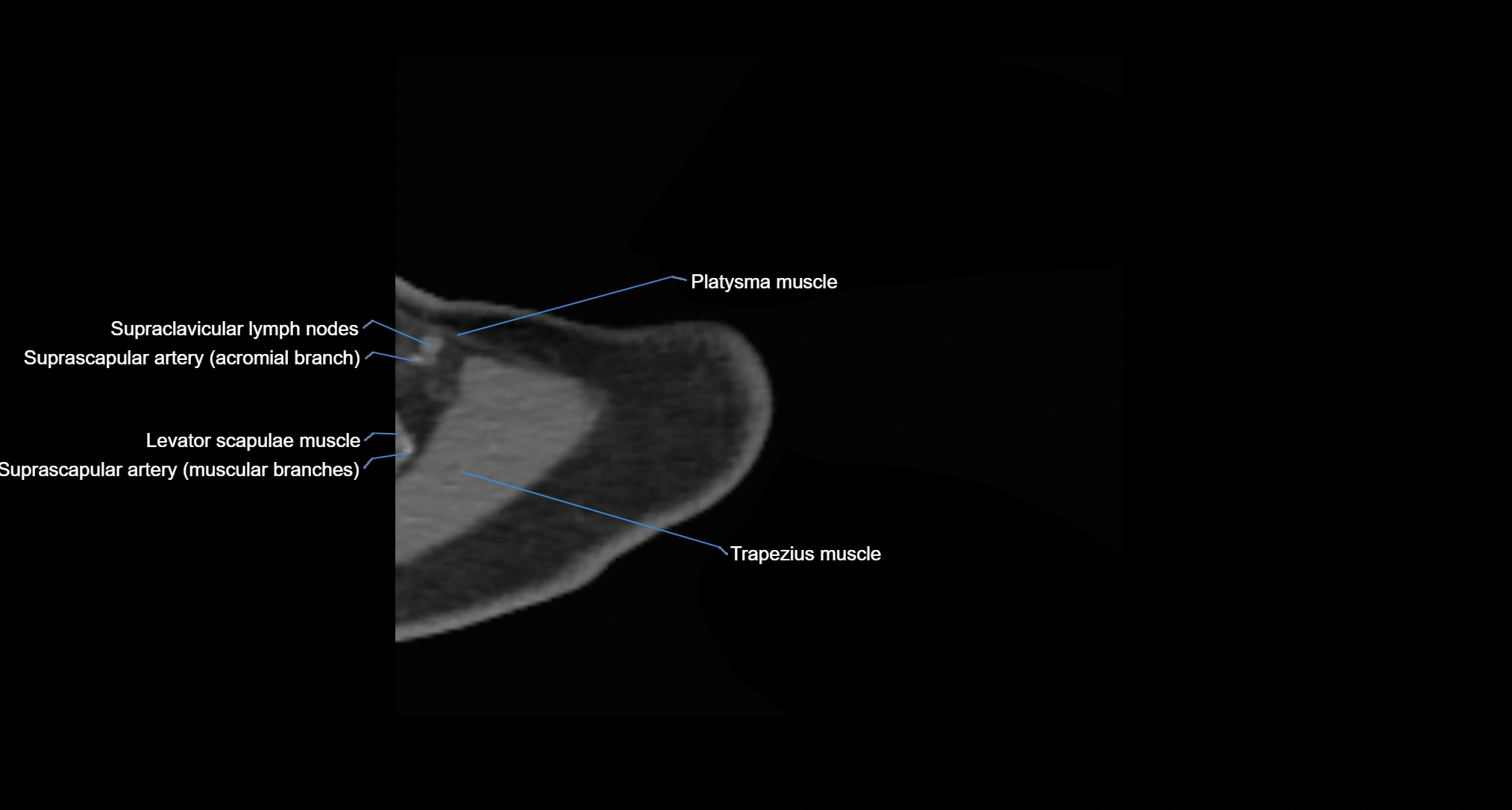
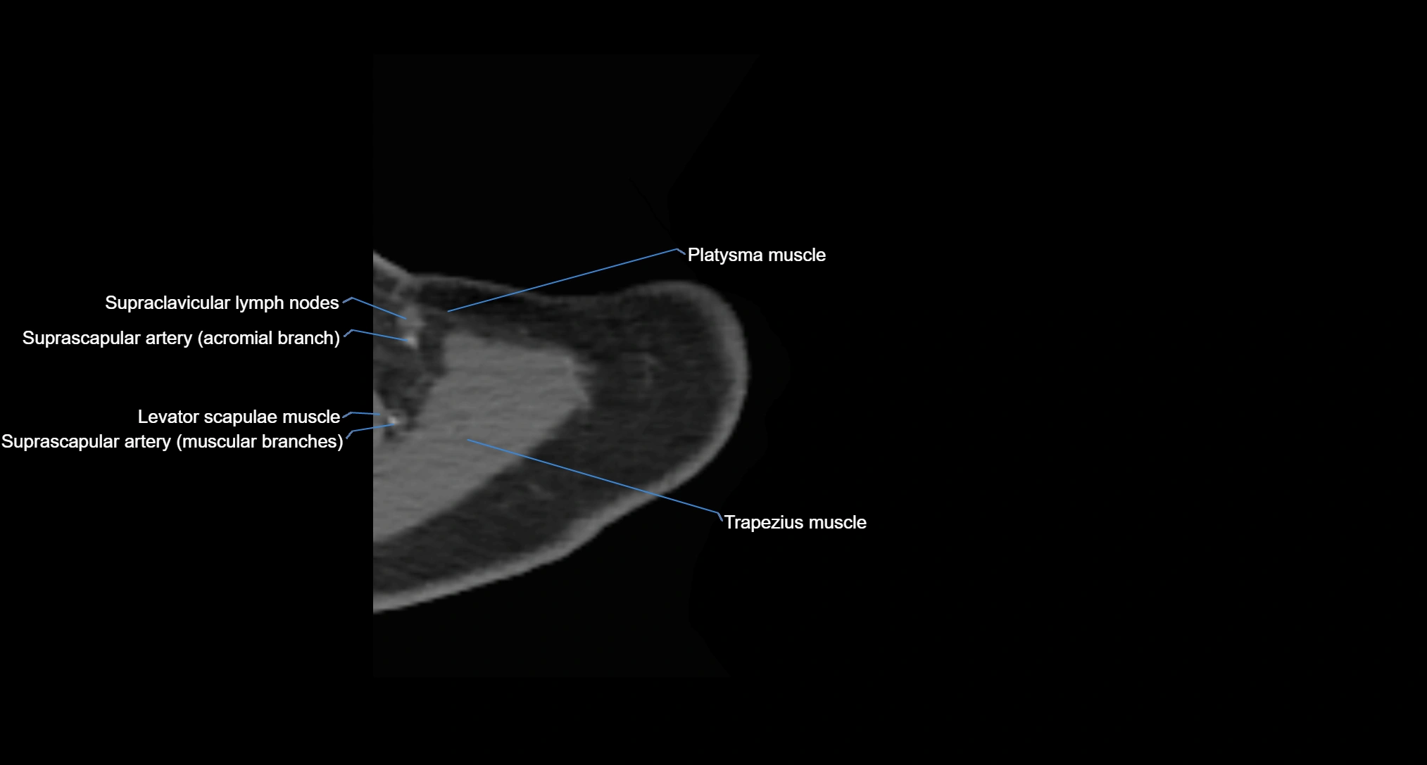
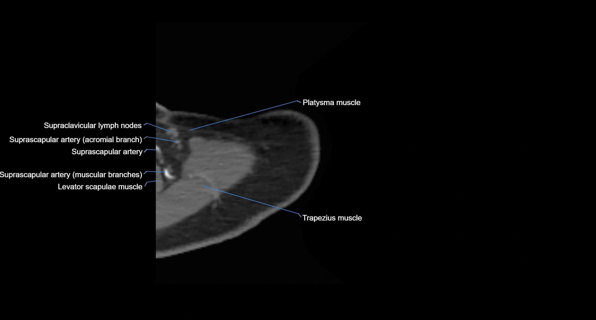
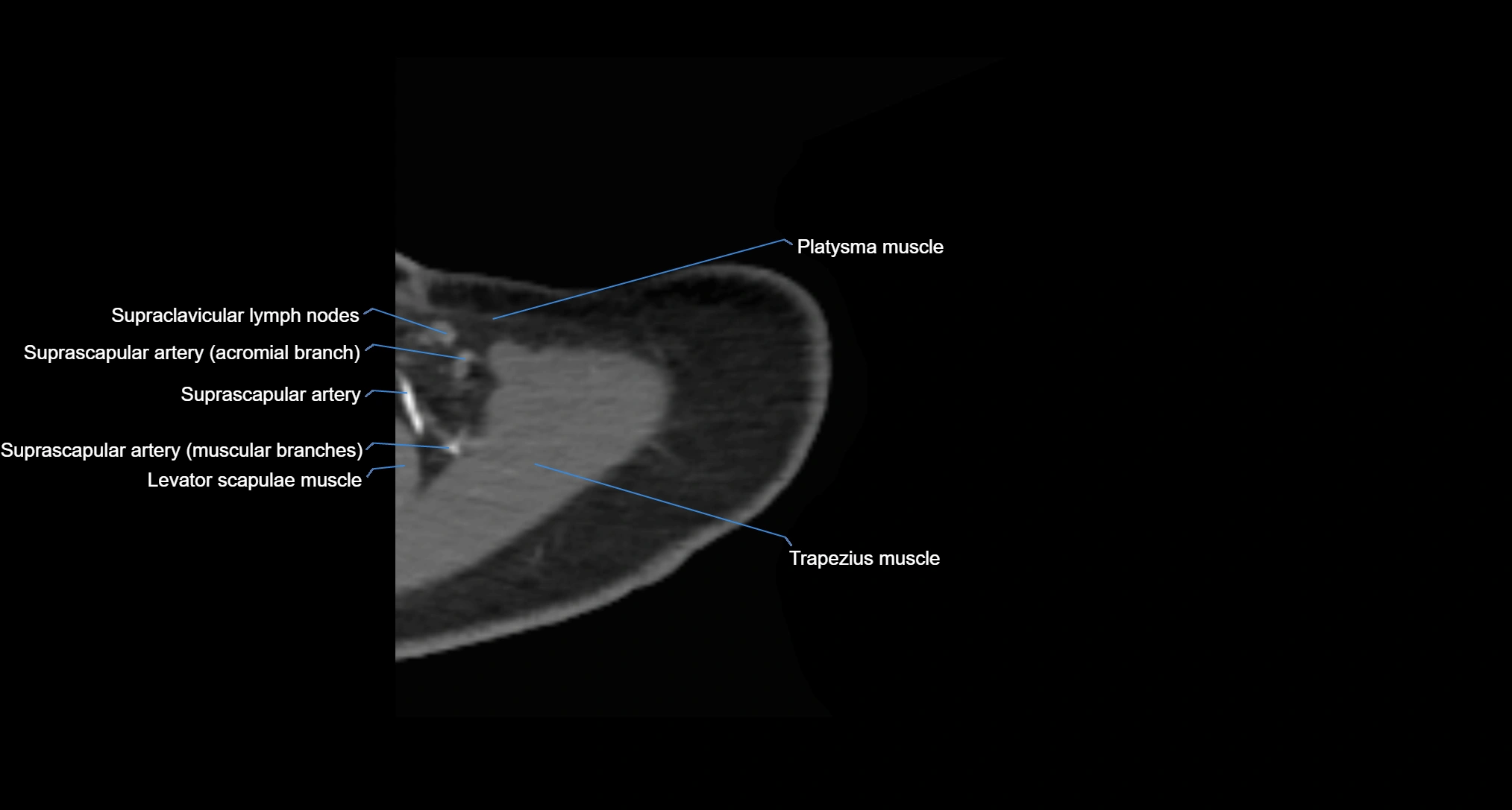
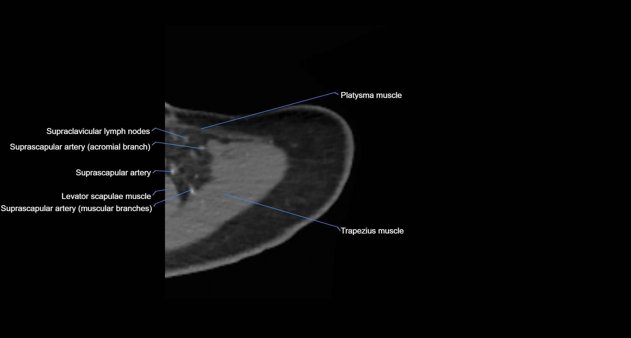
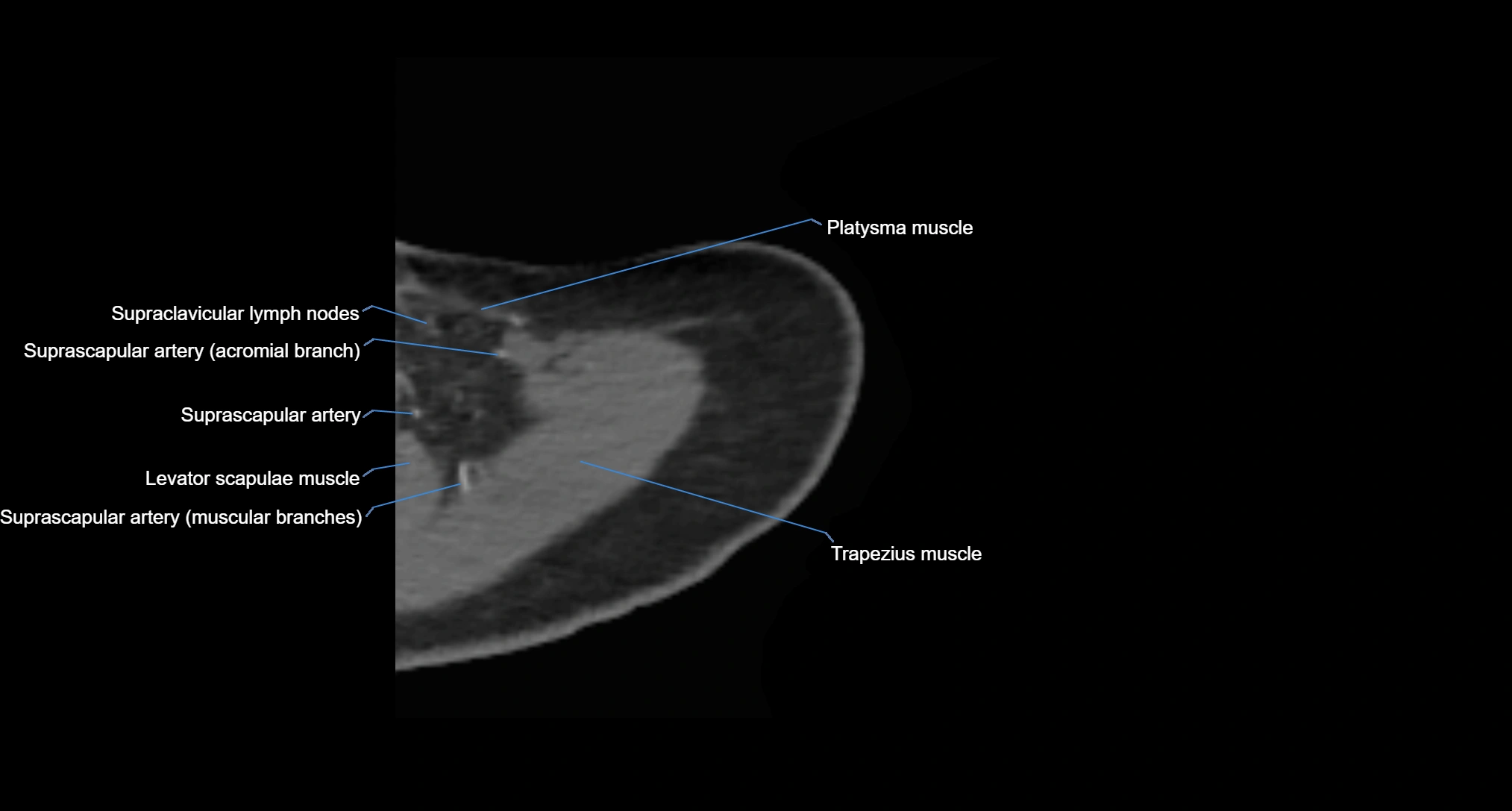
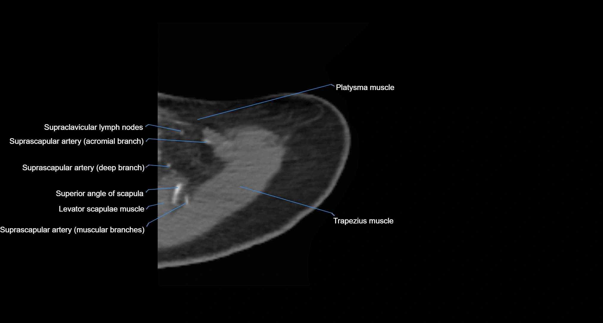
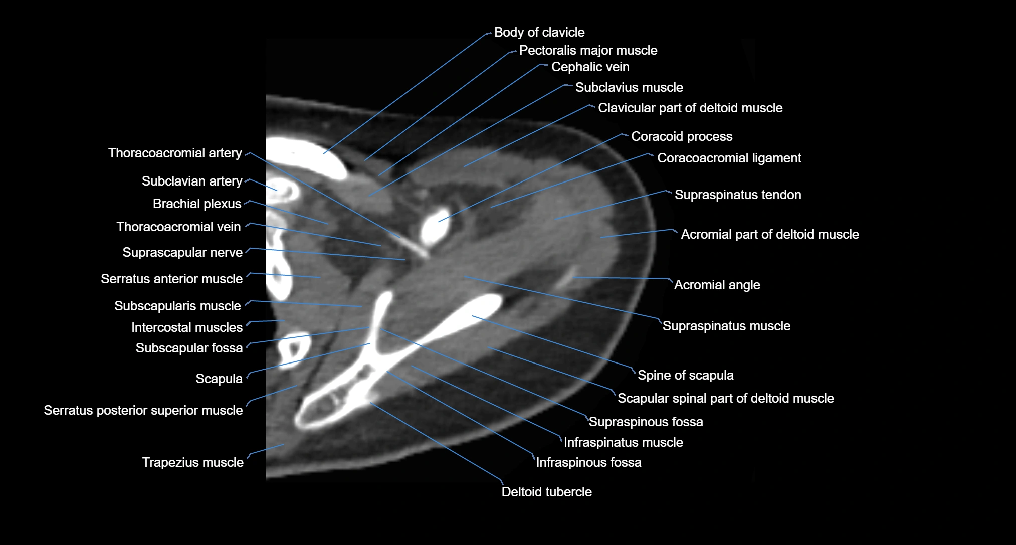
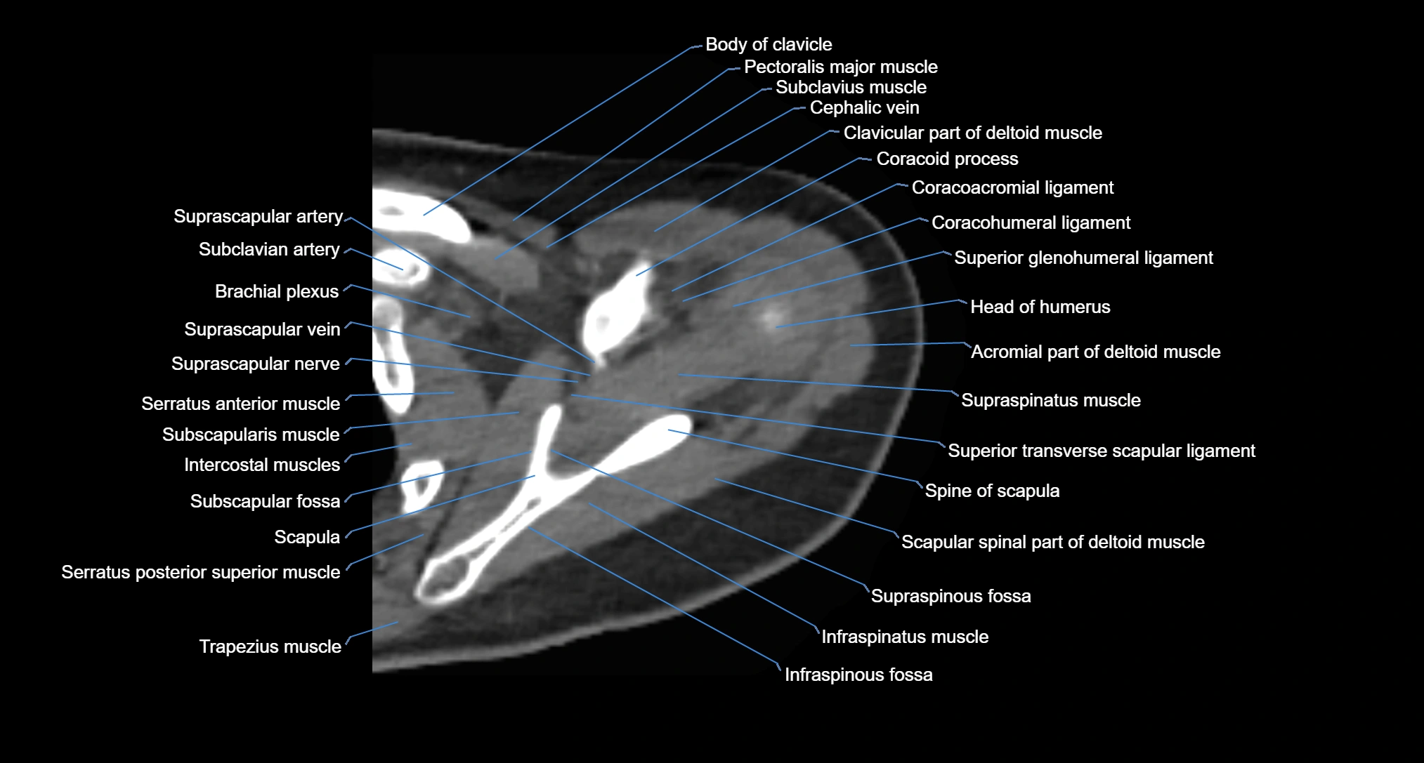
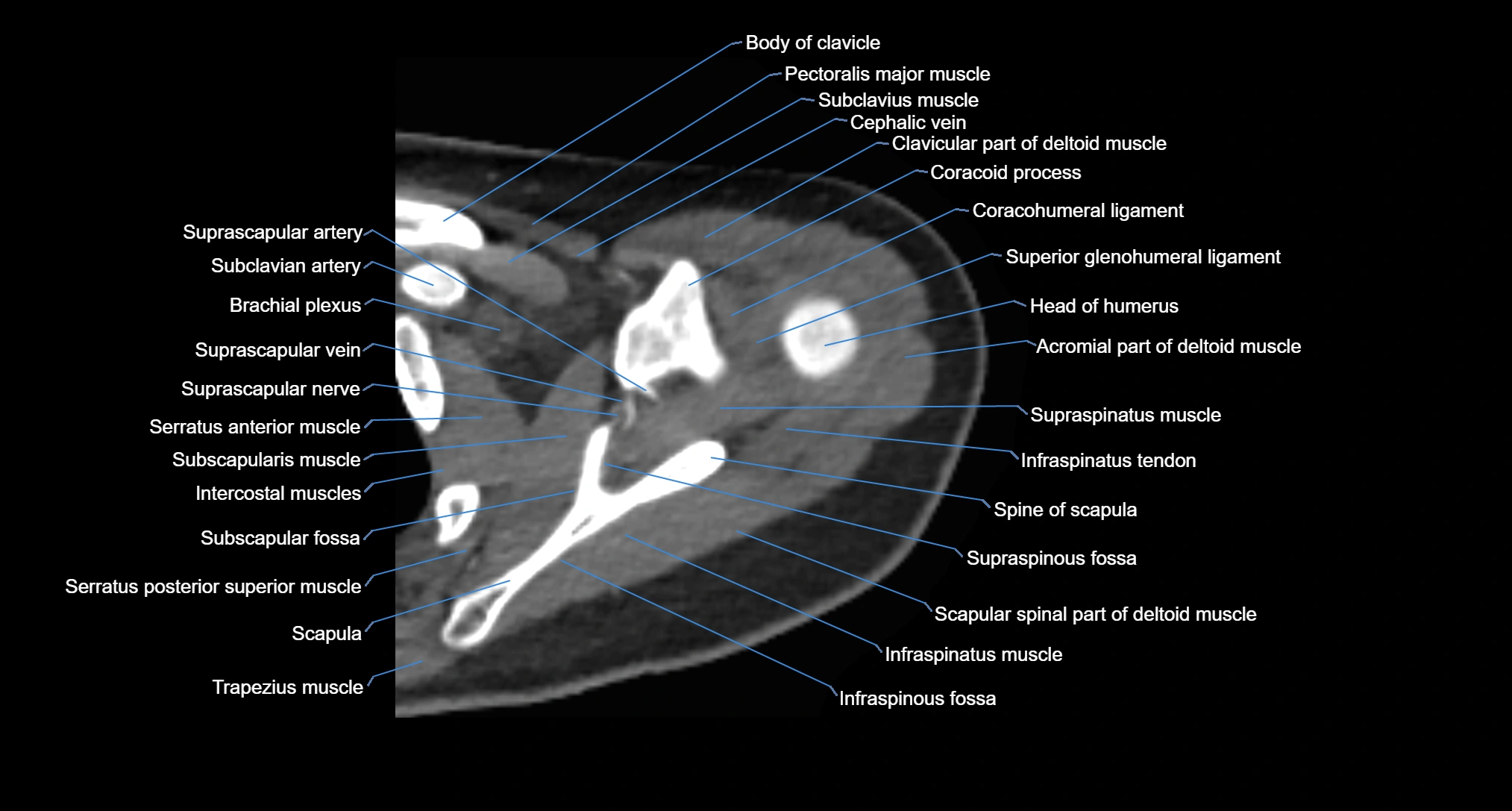
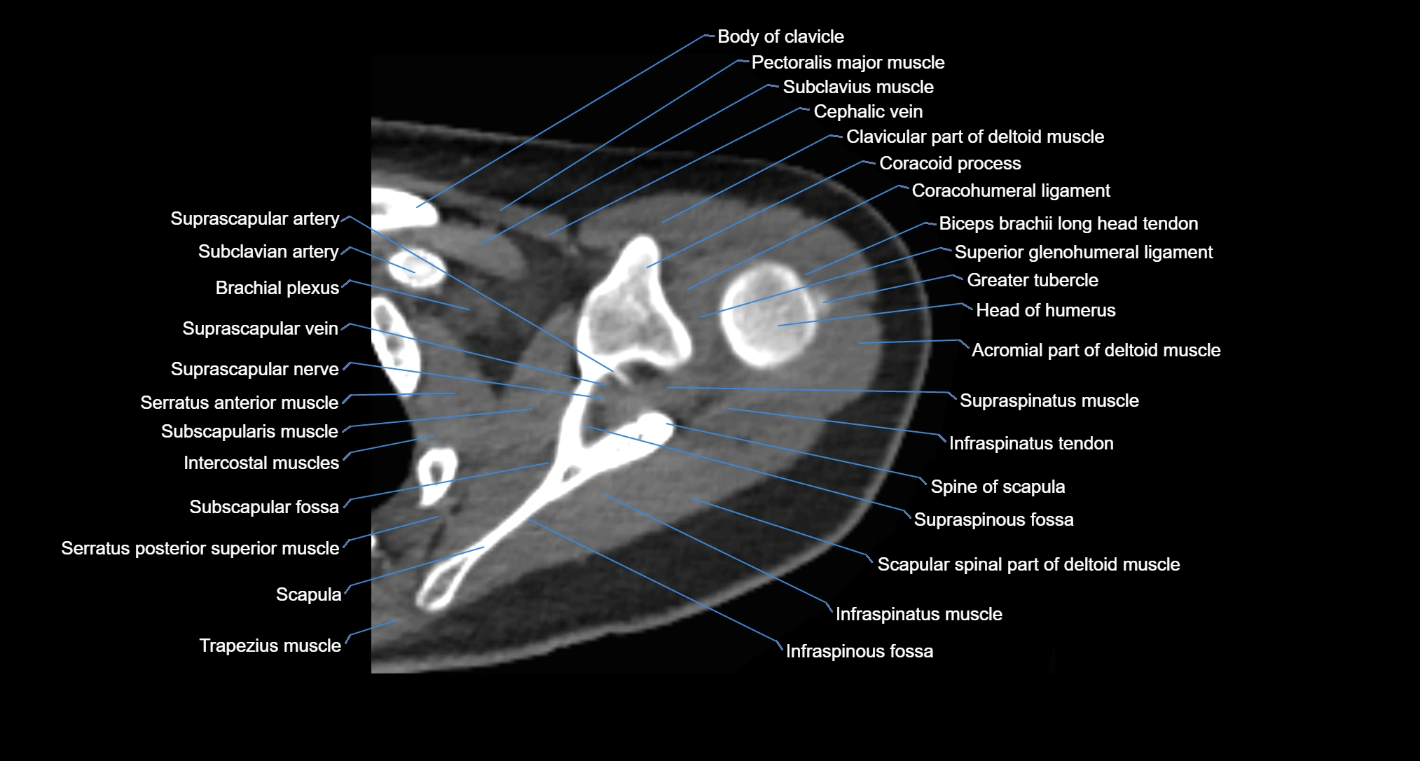
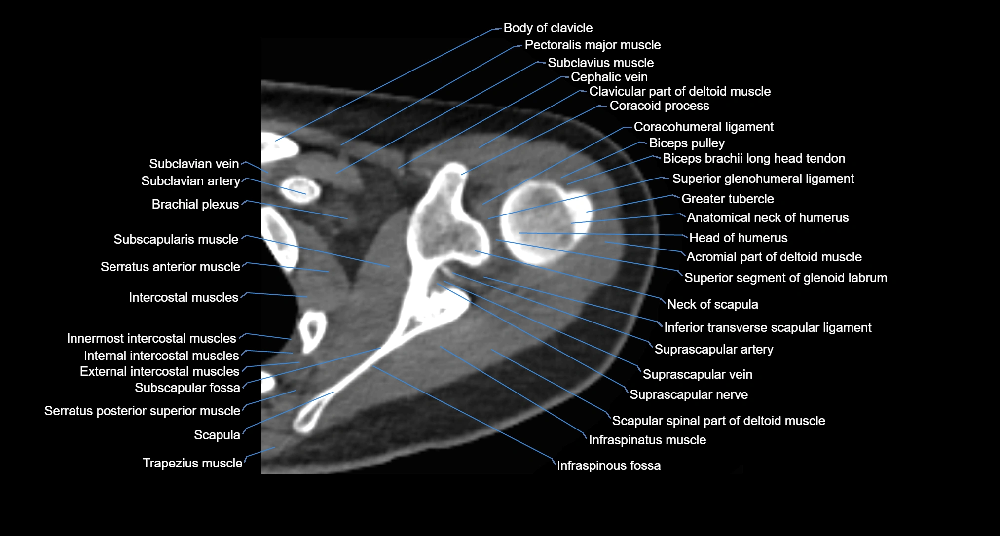
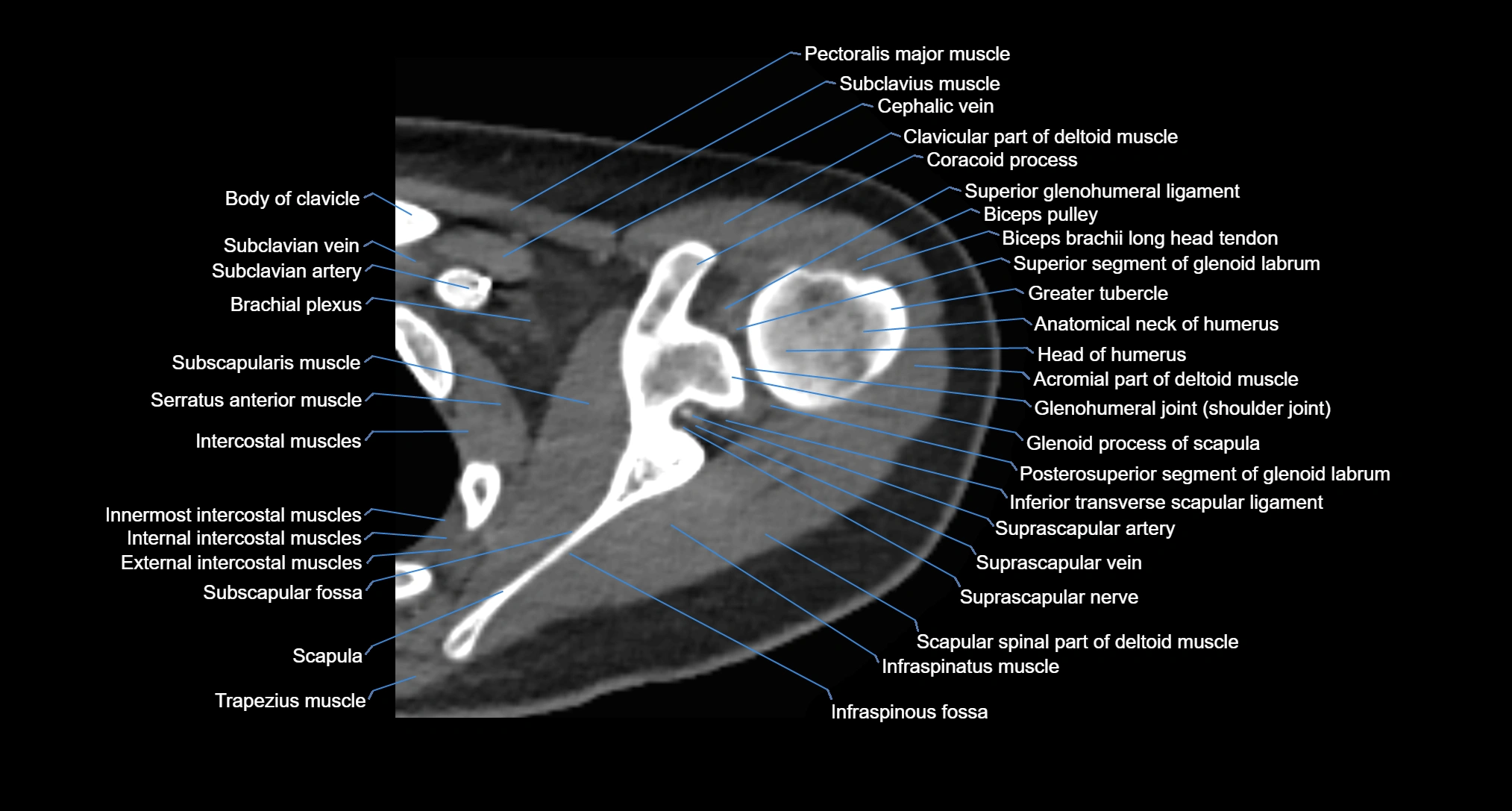
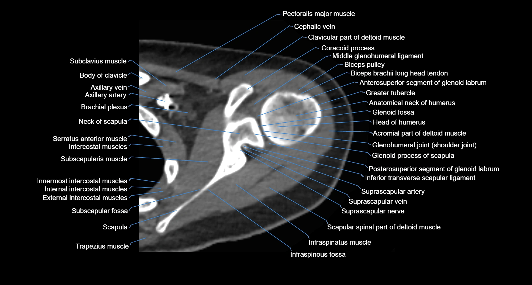
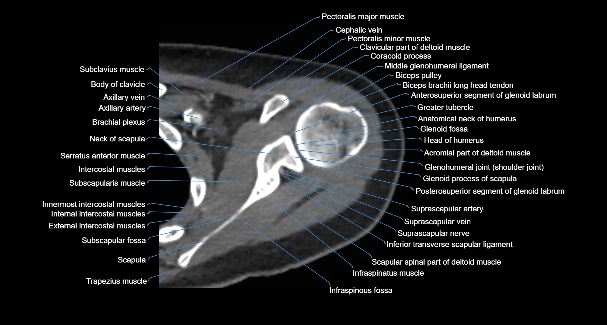
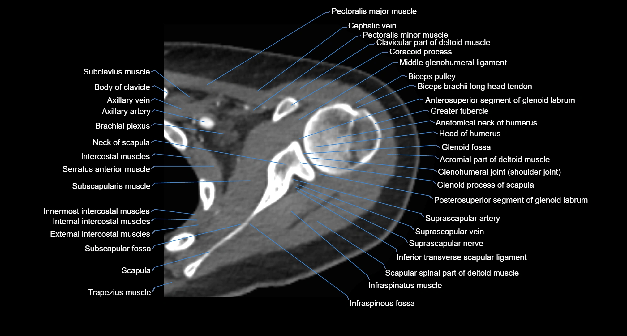
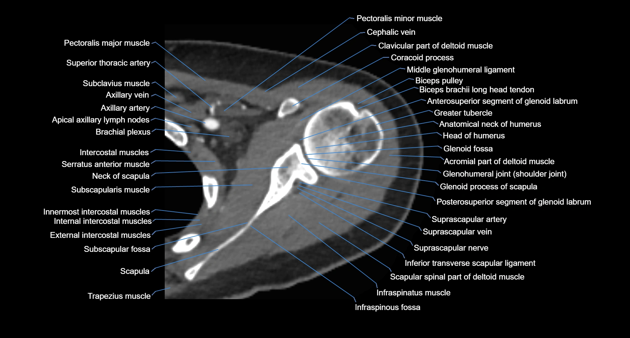
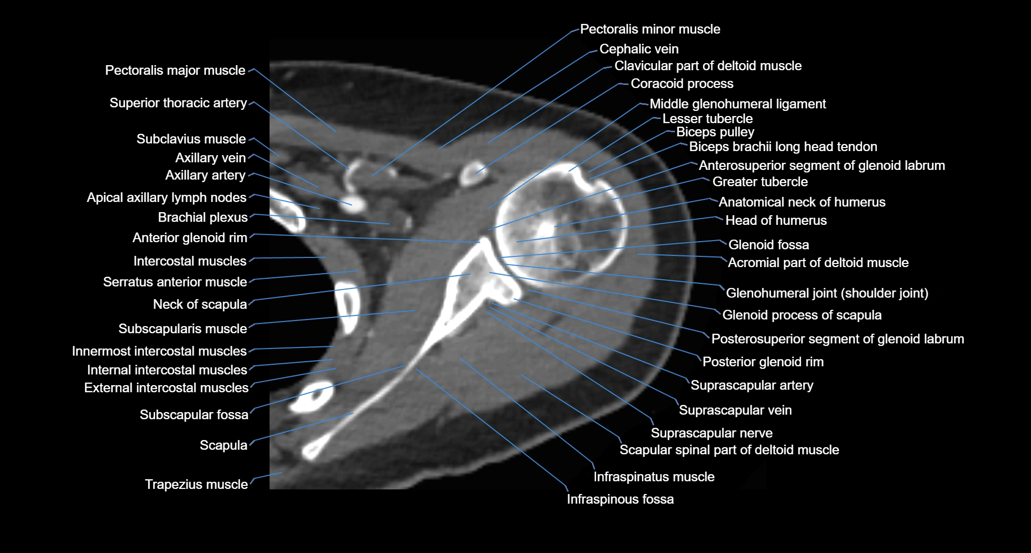
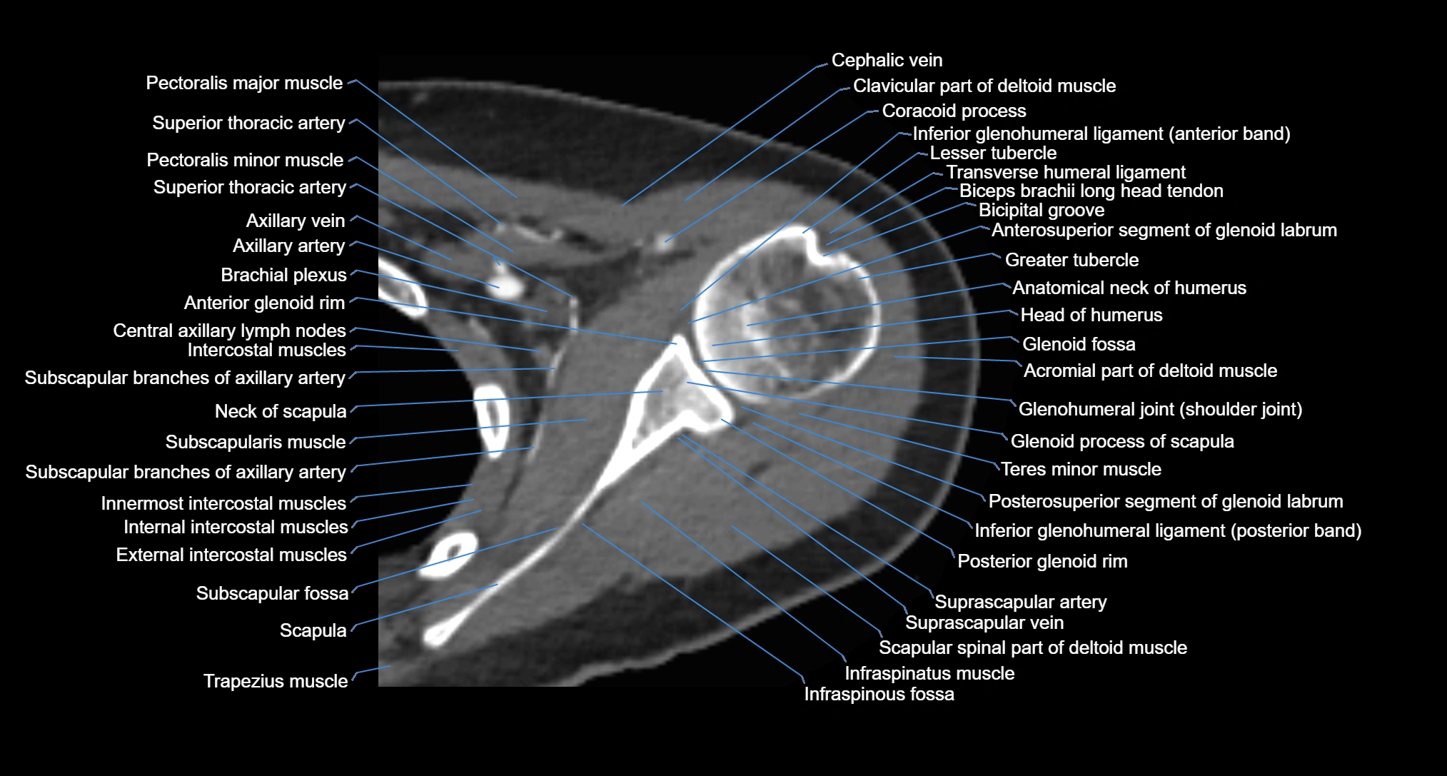
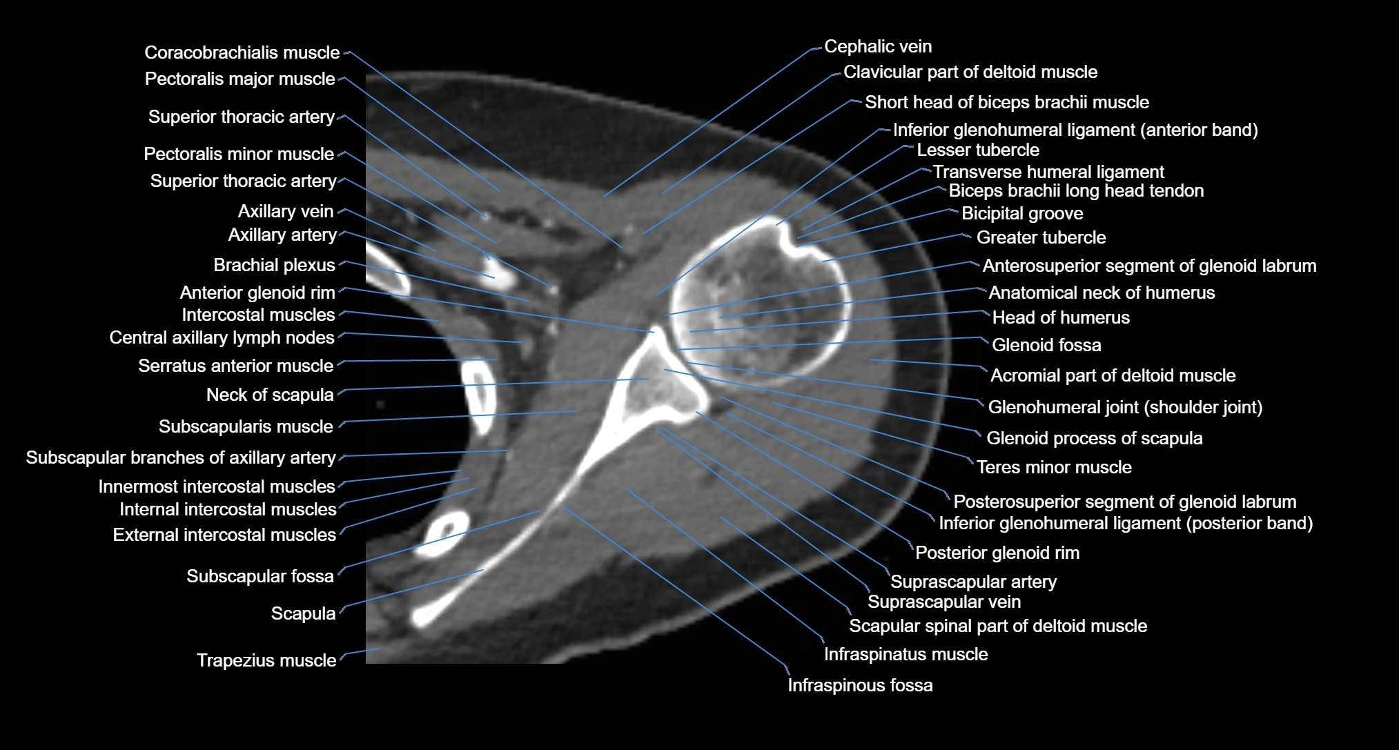
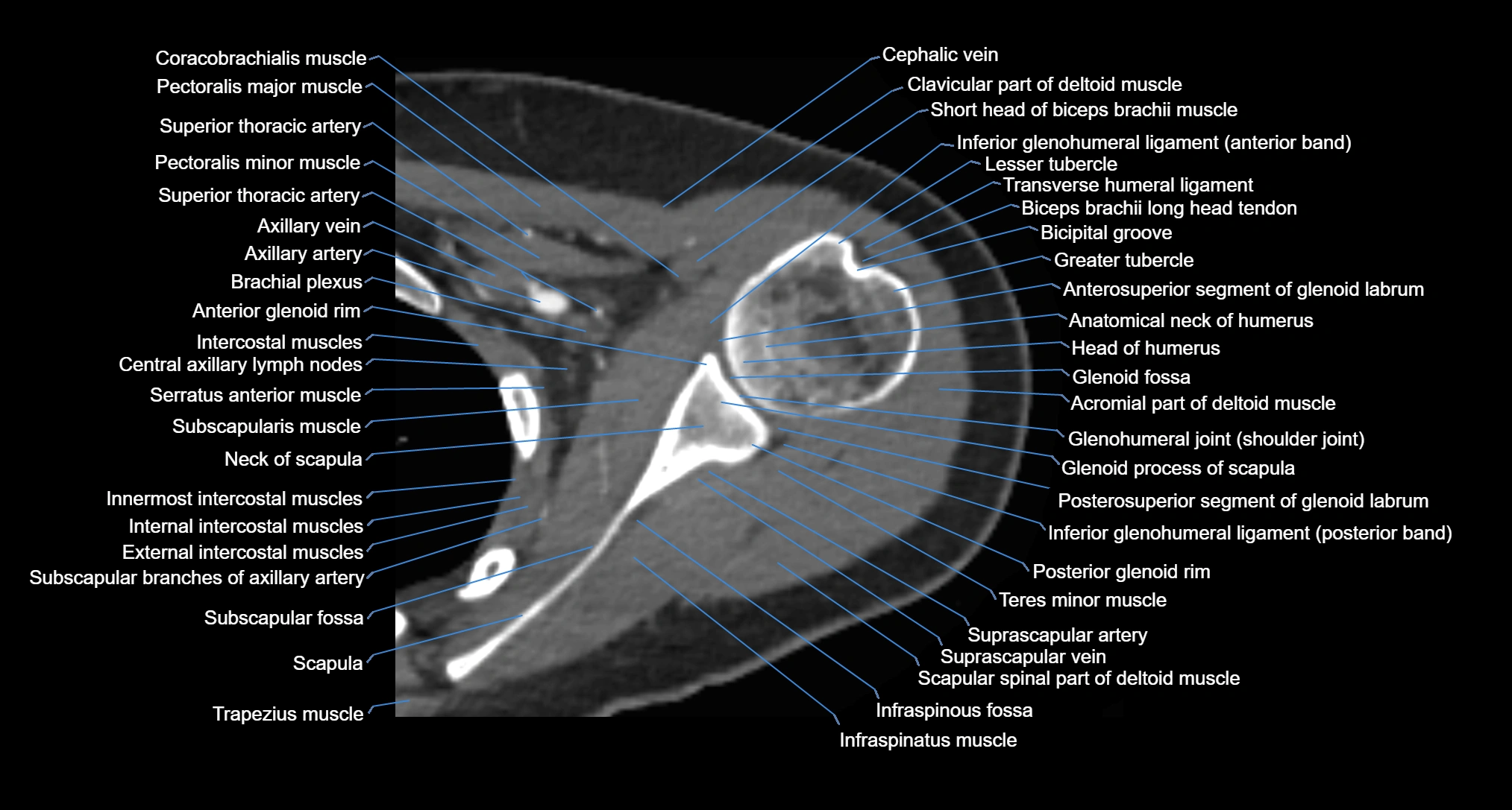
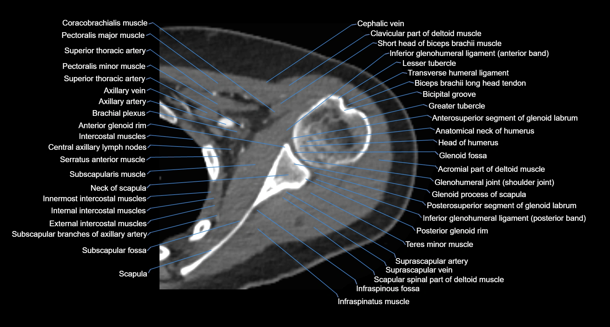
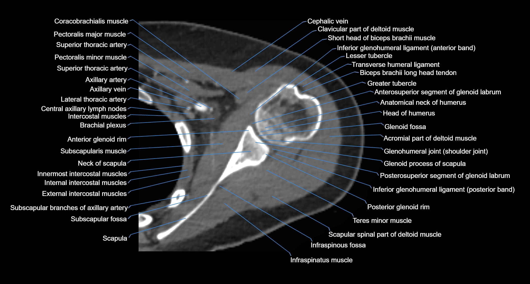
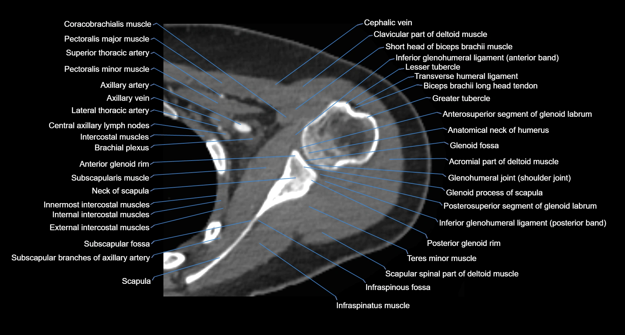
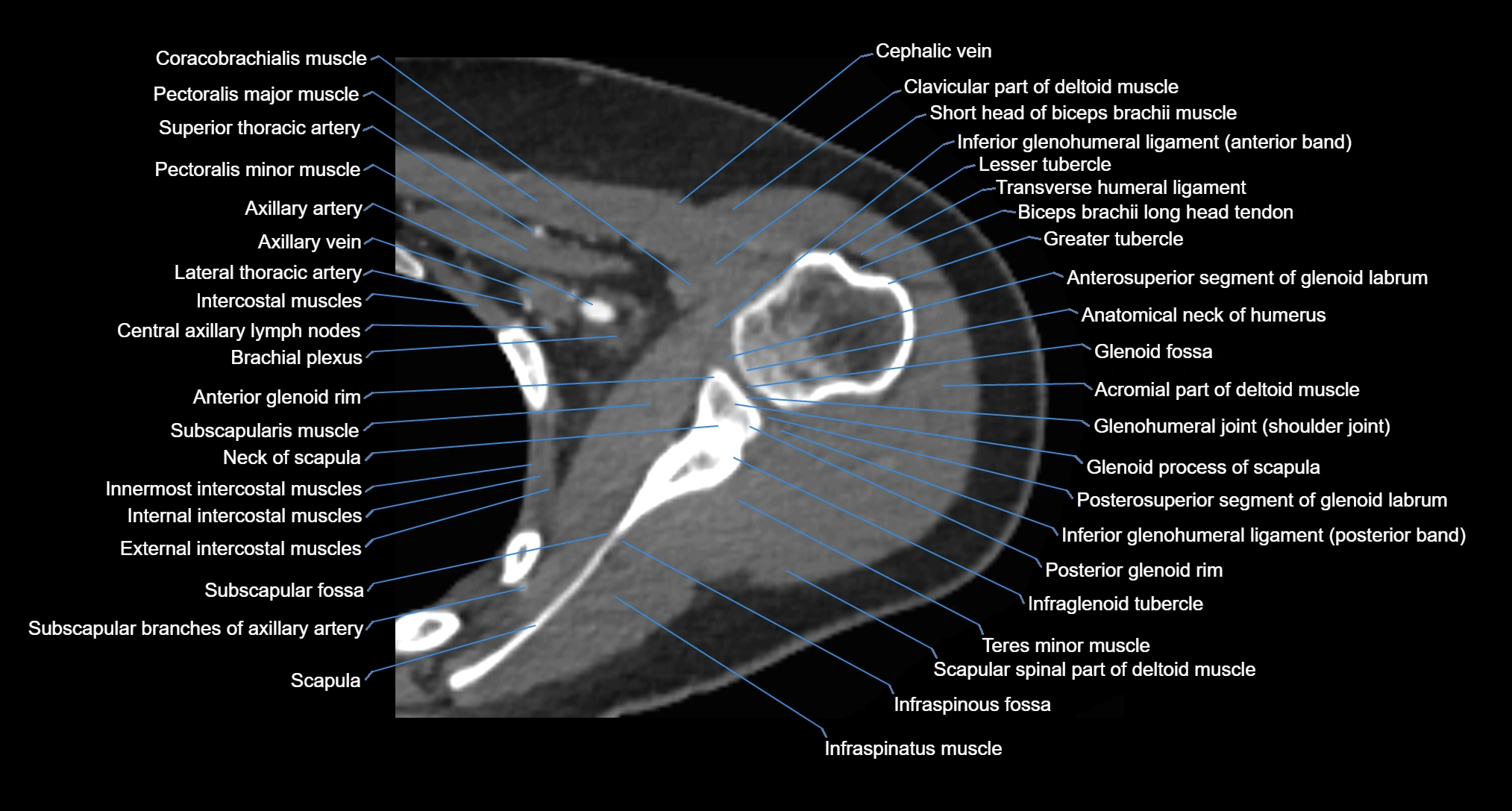
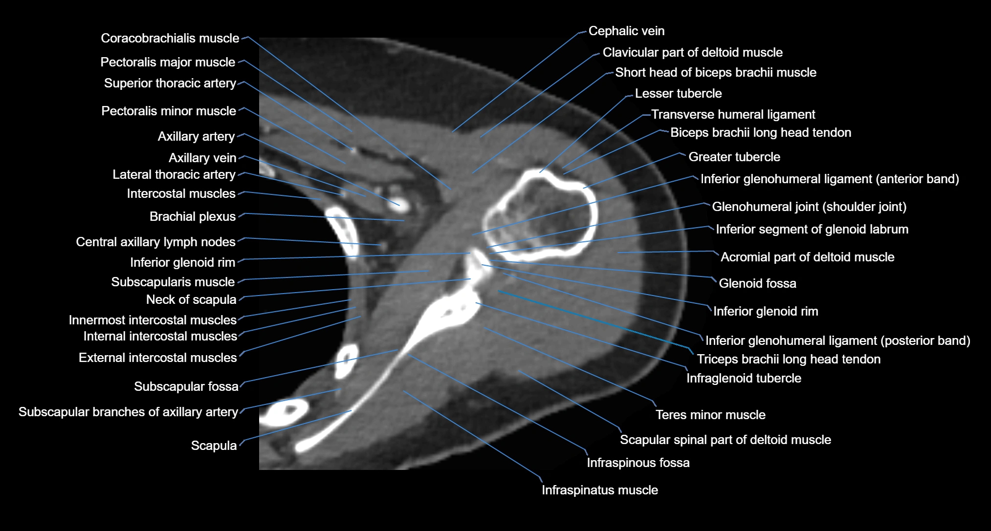
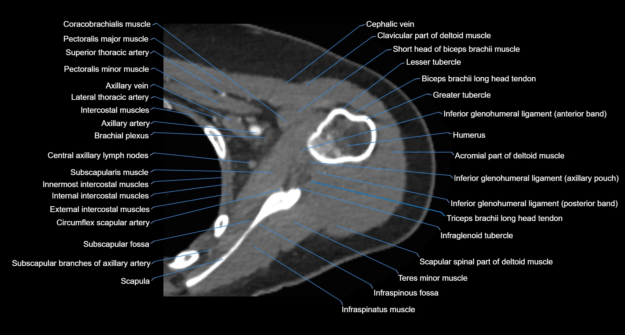
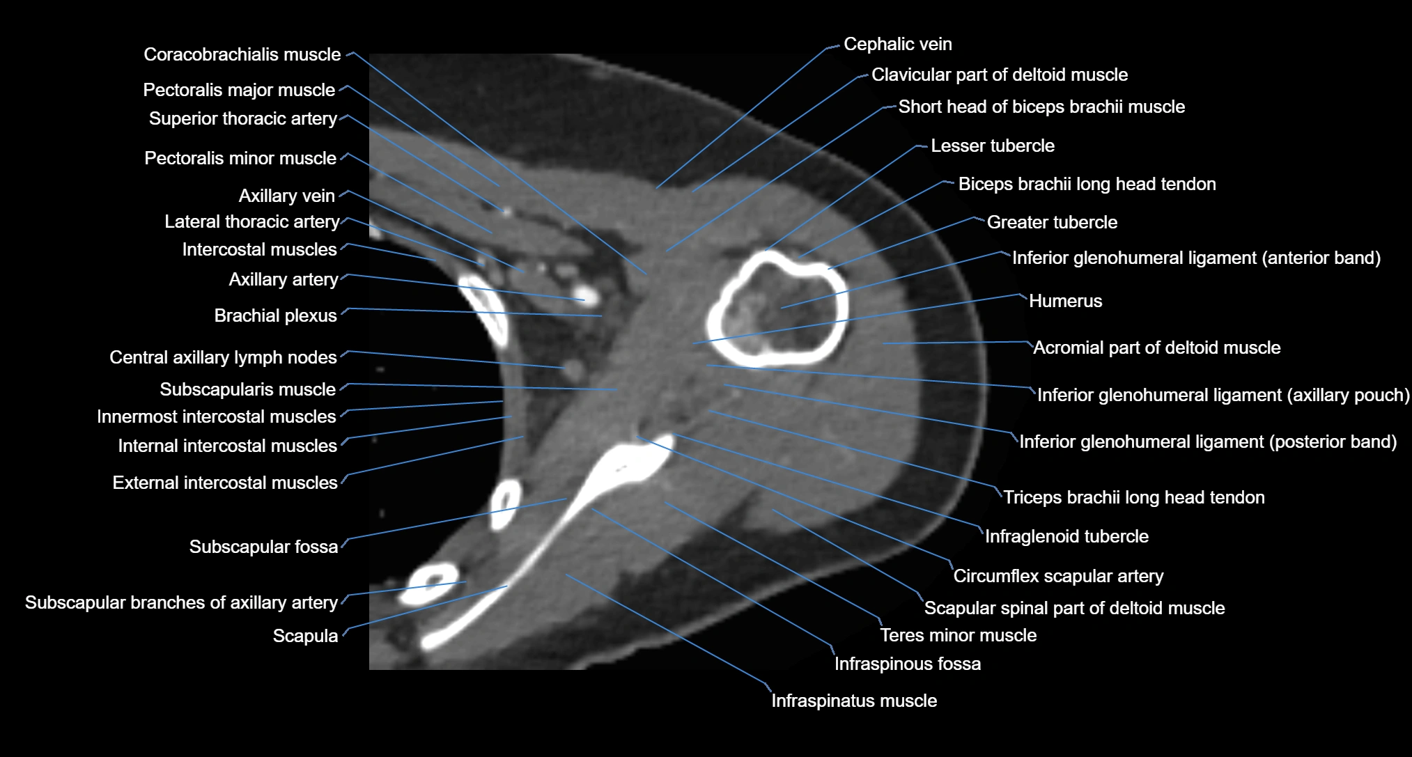
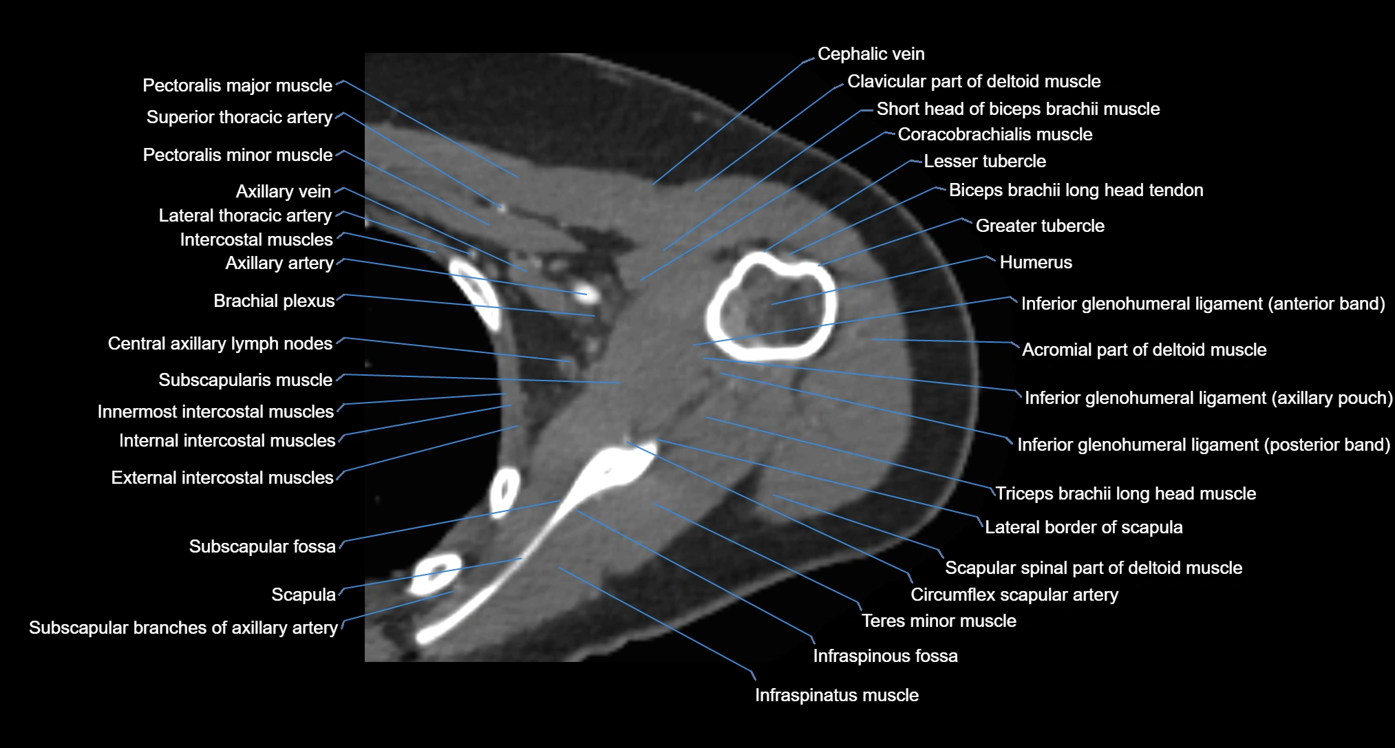
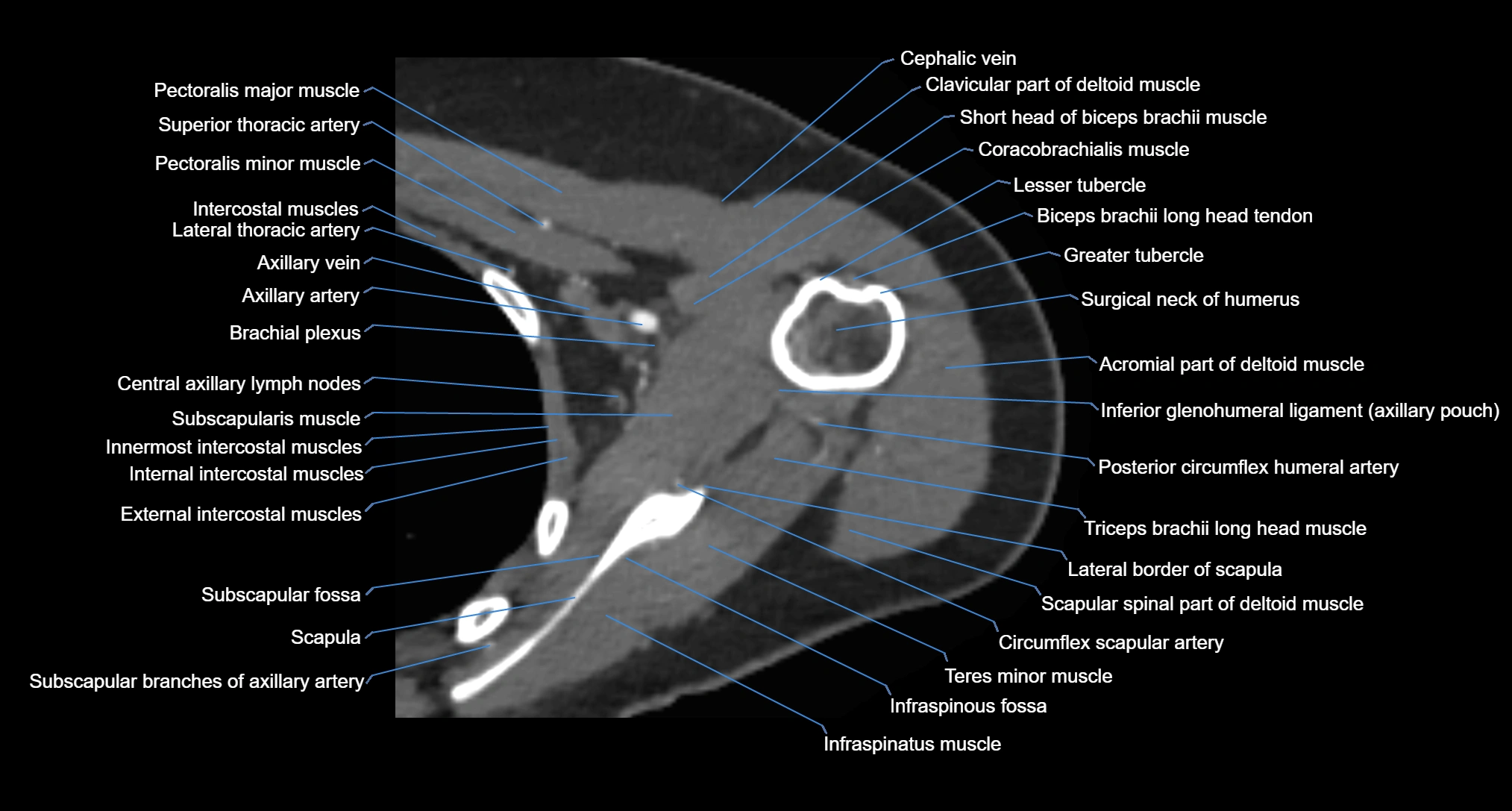
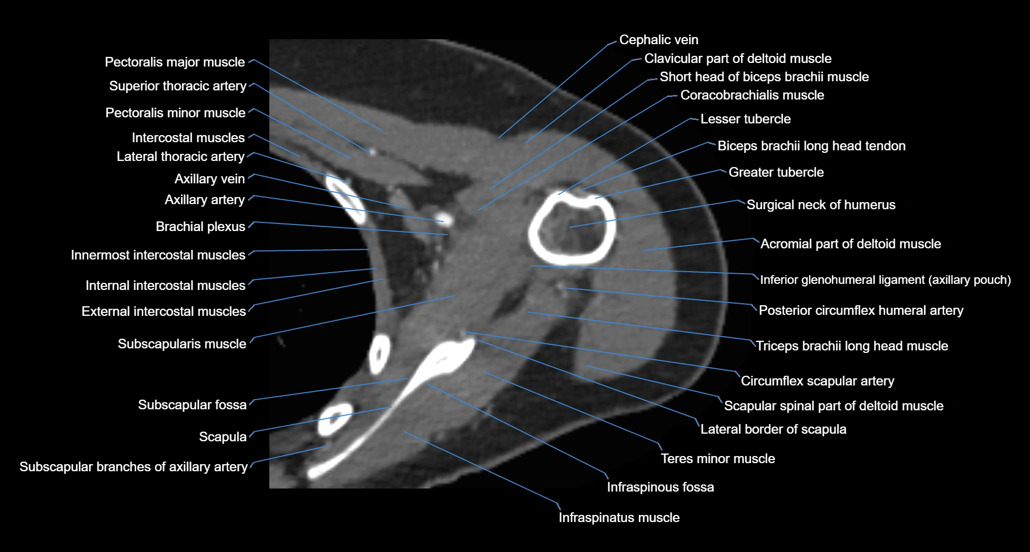
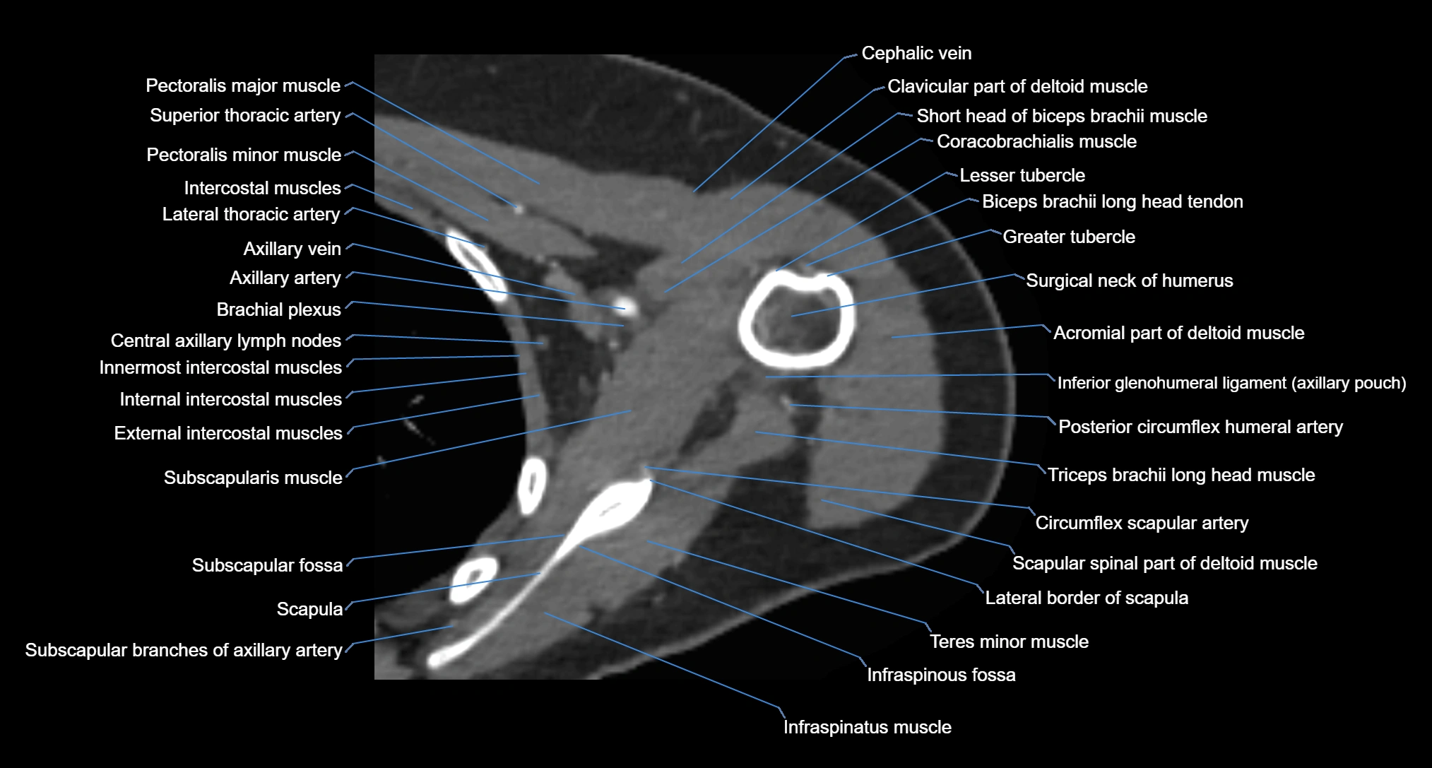
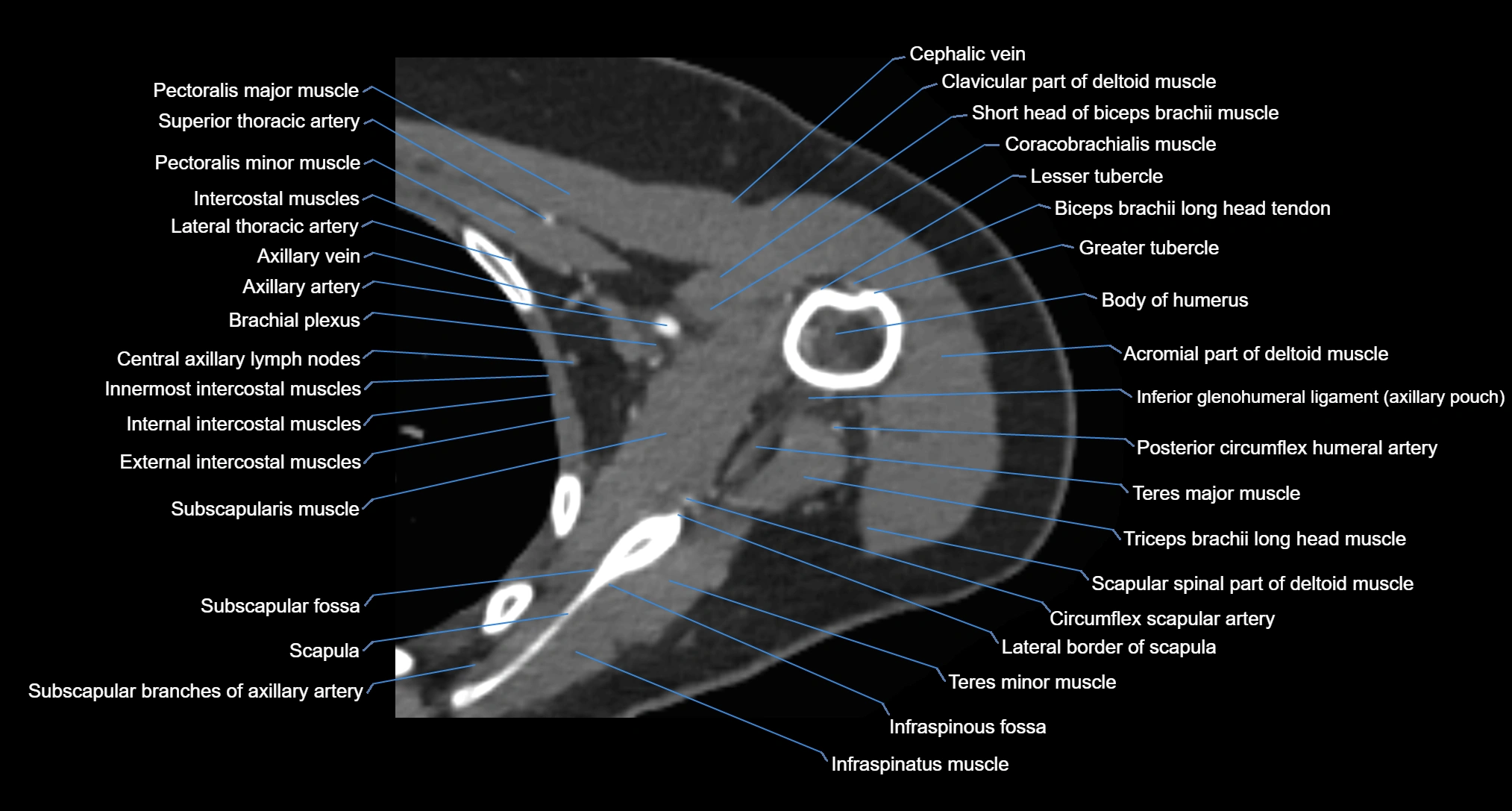
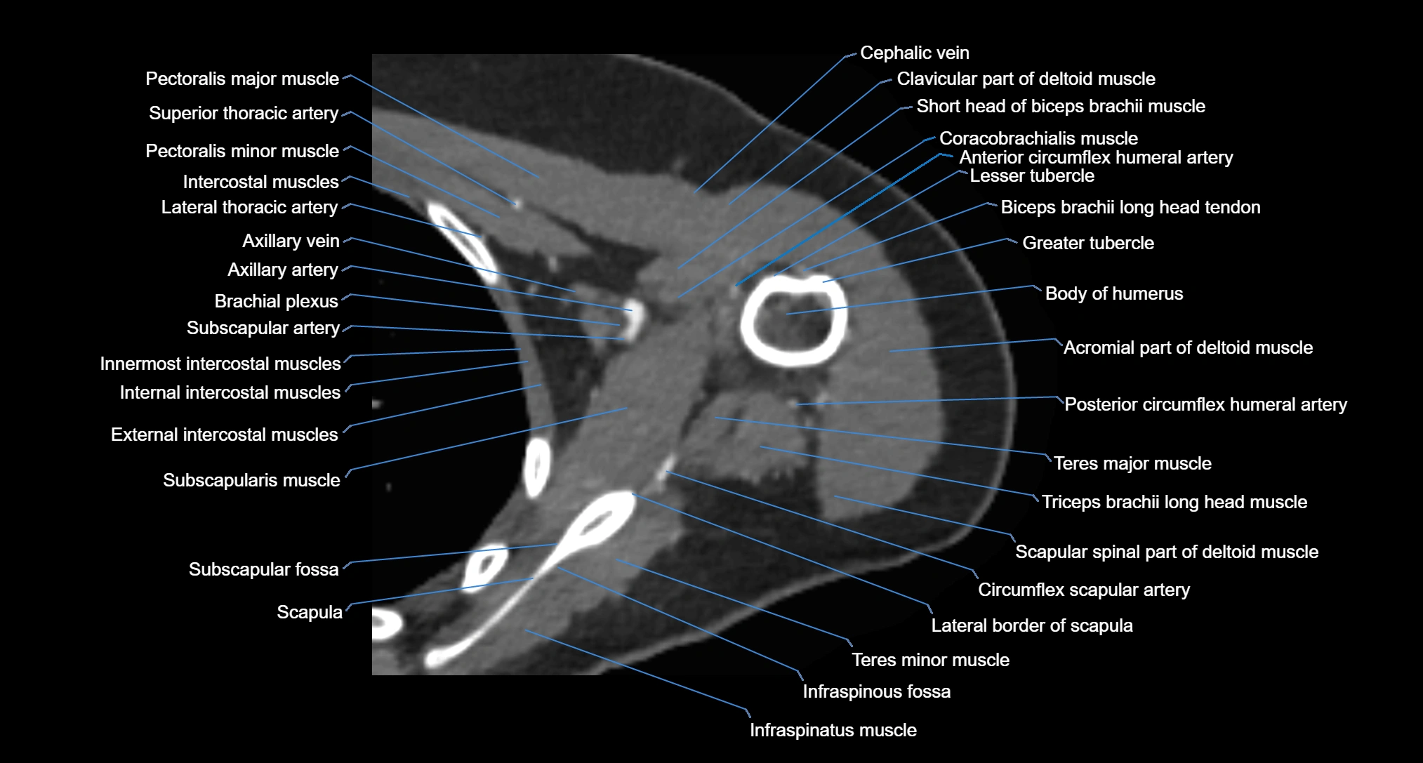
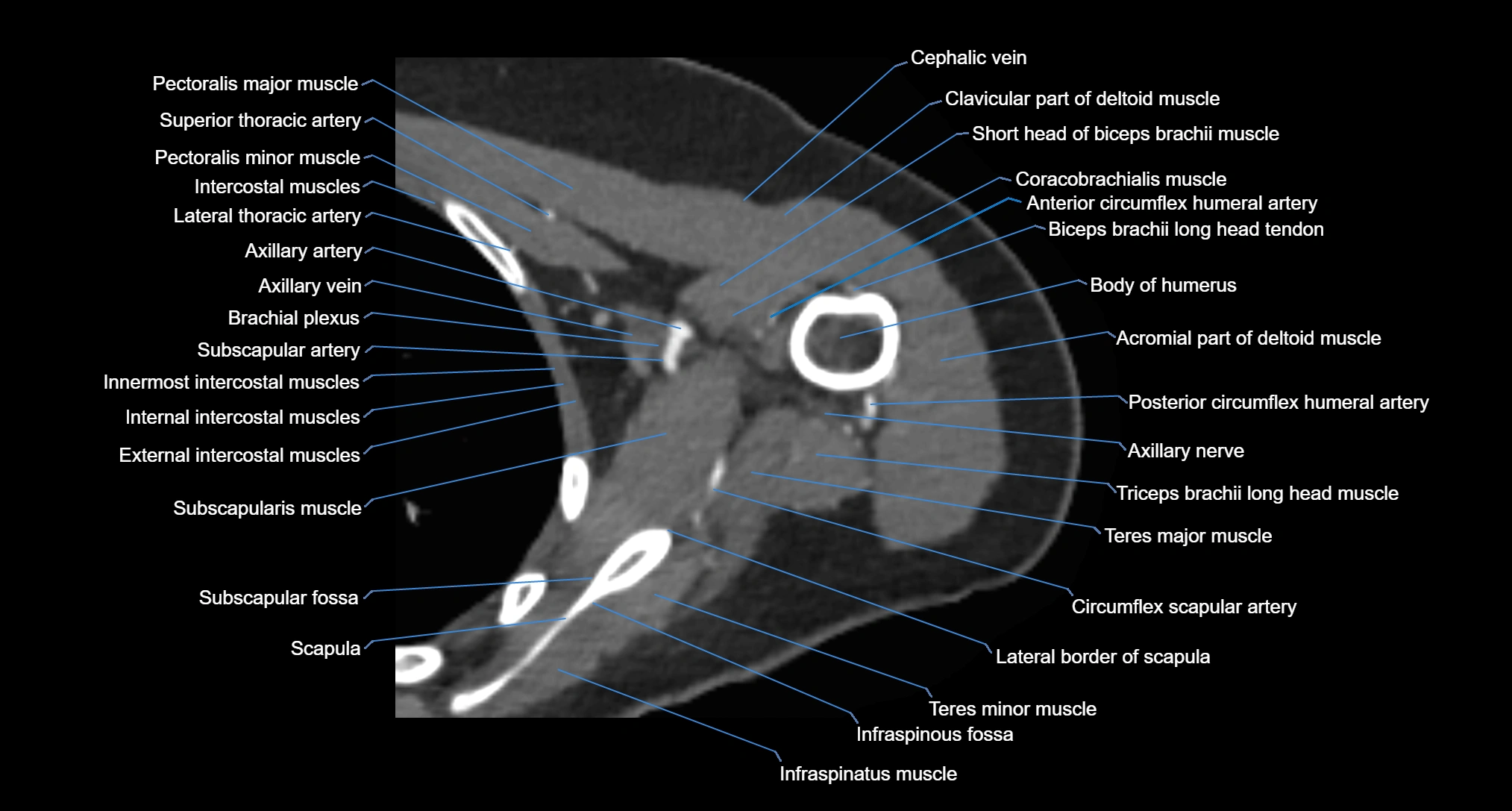
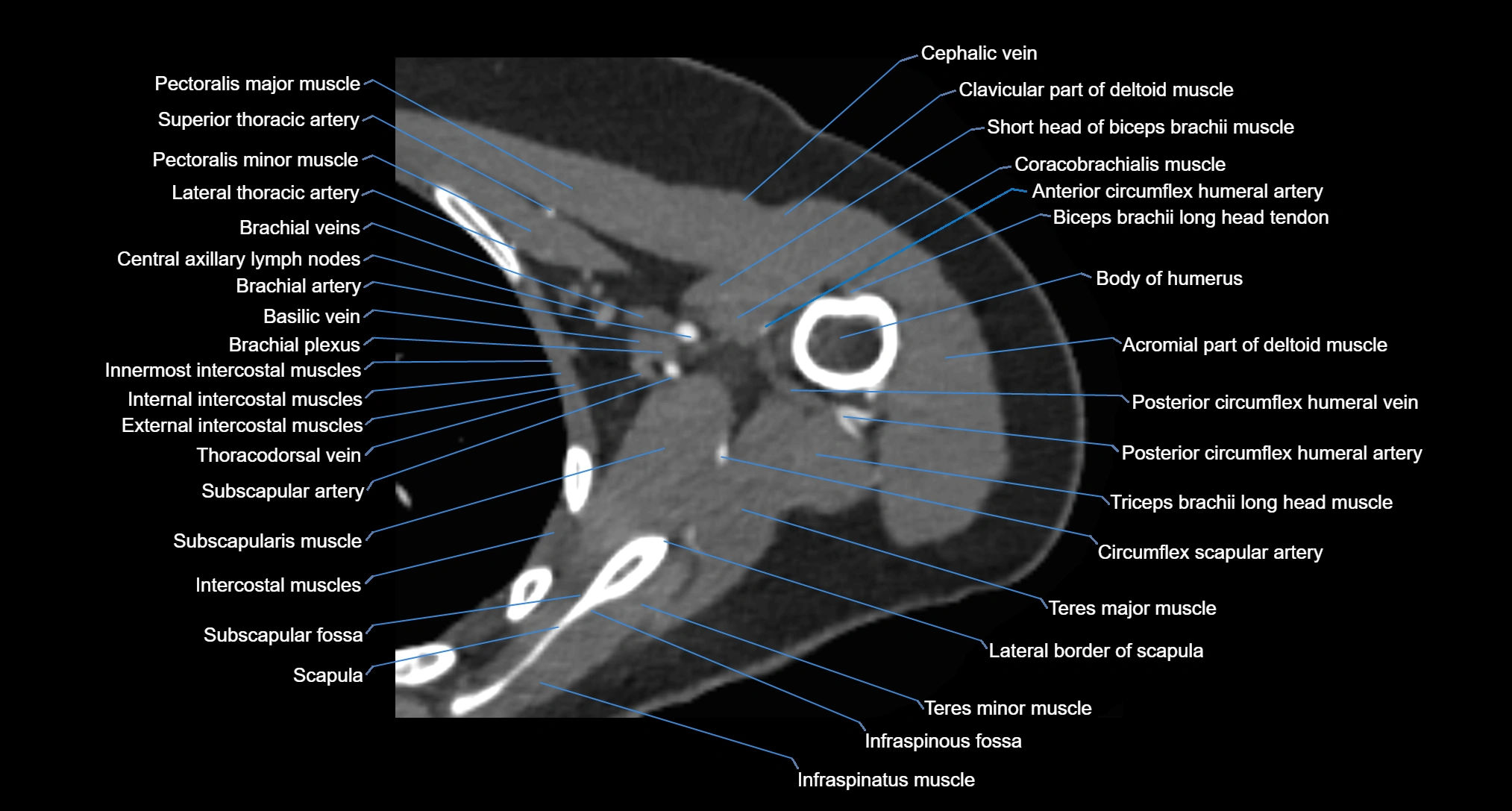
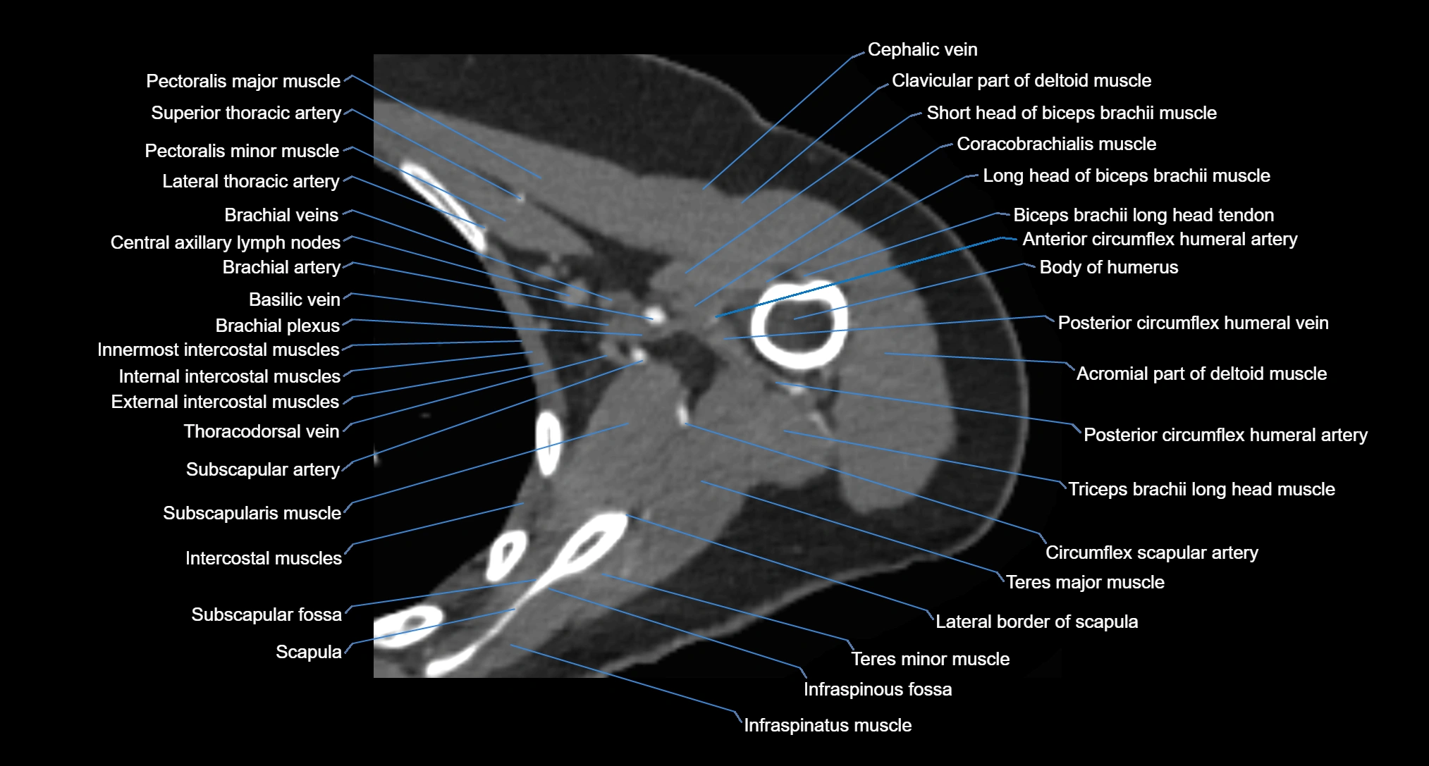
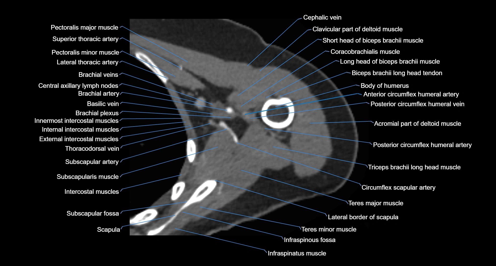
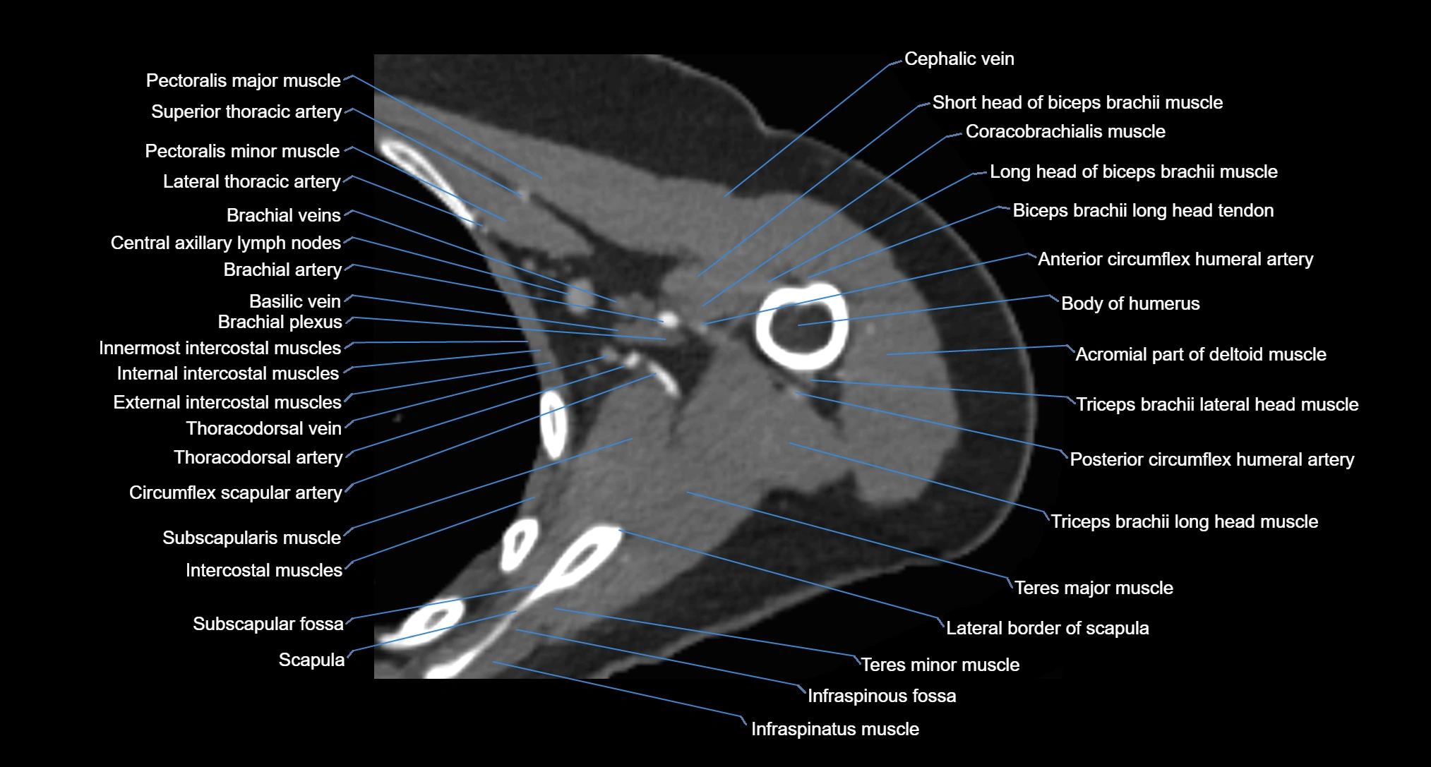
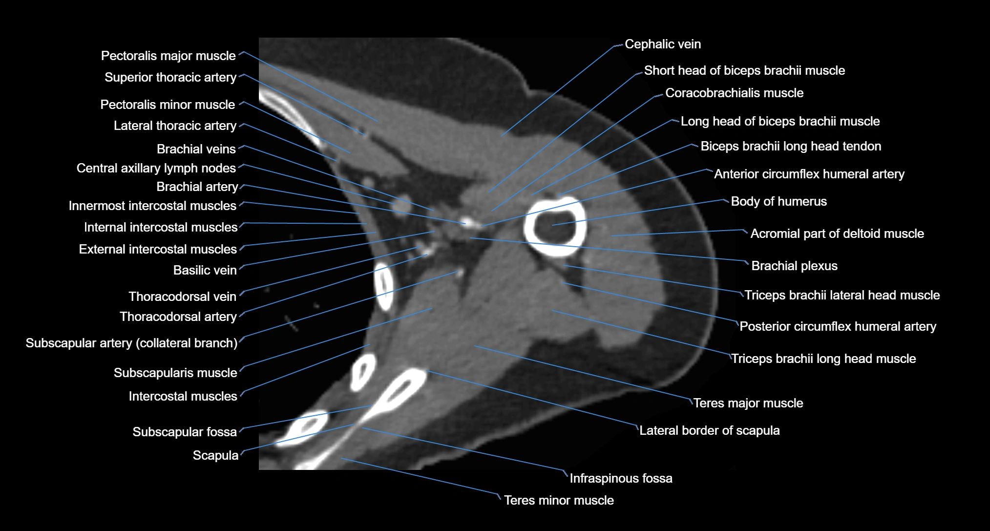
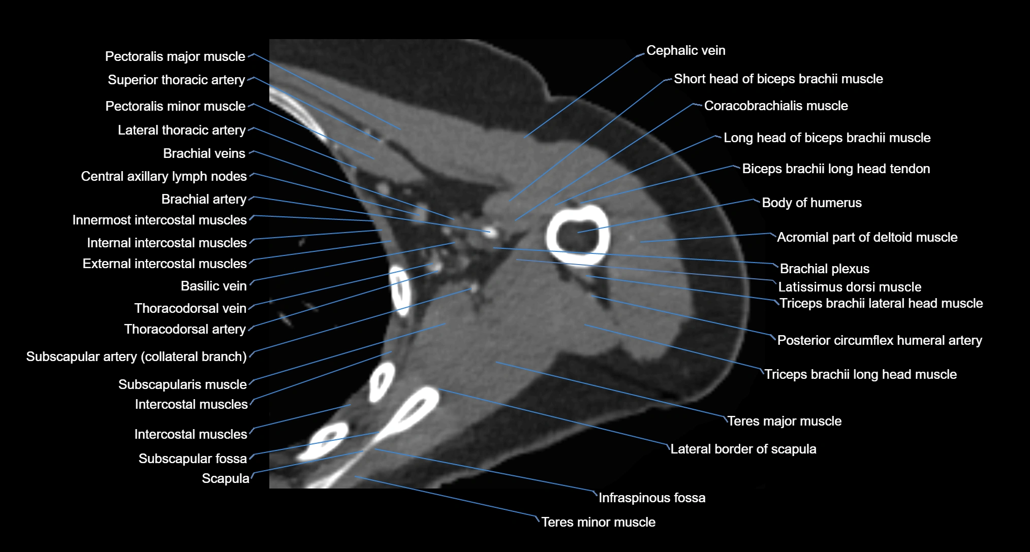
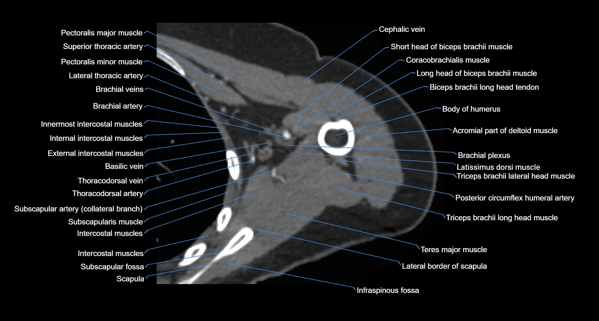
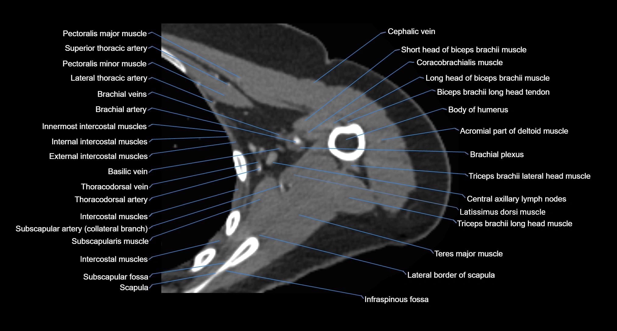
CT image
